Short-Lived Species Might Not Be Much Use in Deciphering the Role of Mitochondrial DNA Damage in Aging

Mitochondria are the power plants of the cell, responsible for producing the energy store molecule ATP that powers cellular operations. Hundreds of these organelles can be found in every cell, the distant descendants of symbiotic bacteria long ago integrated into core cellular mechanisms. They contain their own small remnant genome, and when worn or damaged, they are broken down and recycled by cellular maintenance mechanisms. Mitochondria reproduce by fission like bacteria, but also fuse together at times, and promiscuously swap component parts among one another. Cells can also transfer mitochondria between them. This makes it something of a challenge to track the consequences of mitochondrial damage in aging.

Mitochondrial DNA is more vulnerable and less capable of repair than the nuclear genome deeper inside cells. Some forms of random damage can knock out mitochondrial genes necessary for the most efficient form of energy production. Mitochondria with this particular problem are inefficient, but also somehow more likely to replicate than their peers: they either evade maintenance processes, or perhaps replicate more rapidly. A cell can be quickly taken over by broken mitochondria running inefficient, harmful forms of energy production. The cell becomes dysfunctional and exports damaging, oxidative molecules into the surrounding tissue. The growth in this sort of problem cell is thought to be one of the root causes of aging.

Definitively proving that to be the case is challenging, short of building the necessary technology to repair or prevent mitochondrial DNA damage, such as the allotopic expression methodology advocated by the SENS Research Foundation. There are various forms of mitochondrial mutator mice with artificially high levels of particular types of mitochondrial DNA damage, and these have been used to both make and counter the argument that only deletion mutations are important in aging. Applying that understanding to normally aging mice is a whole other line of work, however. Investigations in normally aging mice have to date been contradictory and inconclusive; it doesn't help that mitochondria are also subject to a range of other, unrelated changes in behavior and activity with age.

So what to make of the current state of research in this part of the field? The two papers I point out here might be taken as the basis for considering that short-lived species simply don't experience this cause of aging to any significant degree. They do not undergo meaningful amounts of stochastic mitochondrial DNA damage. That might go some way towards explaining why earlier investigations have so far not led to the desired destination.

Clonal expansion of mitochondrial DNA deletions is a private mechanism of aging in long-lived animals

Disruption of mitochondrial metabolism and loss of mitochondrial DNA (mtDNA) integrity are widely considered as evolutionarily conserved mechanisms of aging. Human aging is associated with loss in skeletal muscle mass and function (Sarcopenia), contributing significantly to morbidity and mortality. Muscle aging is associated with loss of mtDNA integrity. In humans, clonally expanded mtDNA deletions colocalize with sites of fiber breakage and atrophy in skeletal muscle. mtDNA deletions may therefore play an important, possibly causal role in sarcopenia.

The nematode Caenorhabditis elegans also exhibits age-dependent decline in mitochondrial function and a form of sarcopenia. However, it is unclear if mtDNA deletions play a role in C. elegans aging. Here, we report identification of 266 novel mtDNA deletions in aging nematodes. Analysis of the mtDNA mutation spectrum and quantification of mutation burden indicates that (a) mtDNA deletions in nematode are extremely rare, (b) there is no significant age-dependent increase in mtDNA deletions, and (c) there is little evidence for clonal expansion driving mtDNA deletion dynamics. Thus, mtDNA deletions are unlikely to drive the age-dependent functional decline commonly observed in C. elegans.

Computational modeling of mtDNA dynamics in C. elegans indicates that the lifespan of short-lived animals such as C. elegans is likely too short to allow for significant clonal expansion of mtDNA deletions. Together, these findings suggest that clonal expansion of mtDNA deletions is likely a private mechanism of aging predominantly relevant in long-lived animals such as humans and rhesus monkey and possibly in rodents.

Germline and somatic mtDNA mutations in mouse aging

The accumulation of acquired mitochondrial genome (mtDNA) mutations with aging in somatic cells has been implicated in mitochondrial dysfunction and linked to age-onset diseases in humans. Here, we asked if somatic mtDNA mutations are also associated with aging in the mouse. MtDNA integrity in multiple organs and tissues in young and old (2-34 months) wild type mice was investigated by whole genome sequencing.

Remarkably, no acquired somatic mutations were detected in tested tissues. However, we identified several non-synonymous germline mtDNA variants whose heteroplasmy levels (ratio of normal to mutant mtDNA) increased significantly with aging suggesting clonal expansion of inherited mtDNA mutations. Polg mutator mice, a model for premature aging, exhibited both germline and somatic mtDNA mutations whose numbers and heteroplasmy levels increased significantly with age implicating involvement in premature aging. Our results suggest that, in contrast to humans, acquired somatic mtDNA mutations do not accompany the aging process in wild type mice.

Exercise as a Compensatory Therapy for Parkinson's Disease

The short commentary here reports on an investigation of the benefits of exercise as a compensatory therapy to reduce the impact of Parkinson's disease. Physical activity and physical fitness produce benefits that are on a par with many pharmaceutical and other therapies when it comes to the progression of age-related diseases. This is as much a judgement on the feeble, marginal nature of so much of present day medicine as it is a statement on the merits of exercise. These therapies are marginal because they fail to tackle the root causes of aging. They attempt to influence the downstream, failing state of cellular activity and metabolism. It is akin to changing the oil in an old engine and pressing the accelerator harder rather than replacing the problem parts.

Physical exercise has repeatedly been demonstrated to alleviate comorbidities associated with aging, and to contribute to reducing an individual's risk of developing neurodegenerative conditions such as Parkinson's disease (PD) or Alzheimer's disease (AD). Evidence has accumulated to suggest that exercise can ameliorate many of the symptoms of PD, not only the motor dysfunction, but also some of the non-motor symptoms (NMS), such as cognitive impairment and depression.

In order to decipher the cellular and molecular mechanisms underlying the potential beneficial effects of exercise in PD, it is necessary to employ animal models. The most common models used by the scientific community focus on replicating the motor symptoms of the disease, by applying a chemical lesion in order to cause degeneration of the dopaminergic pathway, which is responsible for controlling movement. However, such models are not useful for examining the NMS, which typically involve several different neurotransmitter pathways and multiple regions of the brain. A recently-developed animal model, involving induction of α-synuclein overexpression in the adult rat brain using adeno-associated viral (AAV) vectors, is widely considered to most consistently reproduce the pathological features and progressive neurodegeneration associated with human PD.

Adult male rats were given free access to running wheels in cages (voluntary exercise) from one week after administration of AAV-α-synuclein into the substantia nigra. We found that voluntary exercise had no effect on motor function or on dopaminergic neuronal loss in the substantia nigra. However, overexpression of α-synuclein significantly impaired the ability of the animals to perform hippocampal-associated cognitive tasks. This was associated with deficits in hippocampal neurogenesis, a form of neuroplasticity and a key cellular process underlying learning and memory. Importantly, voluntary exercise protected against this cognitive dysfunction, and this protective effect was mediated, at least in part, by alterations in neurogenesis levels.

This is the first study to date that has employed the AAV-α-synuclein model to investigate exercise as a therapeutic intervention, and its strength lies in the fact that this model is widely accepted to be the most similar to the progressive nature of the human condition. It must be appreciated that there are difficulties associated with measuring the effects of exercise in patients, as well as in animal models, that have problems with their motor function. Nevertheless, all of the available evidence suggests a growing rationale for including structured exercise programs as part of a patient's therapeutic regimen.

Link: https://doi.org/10.18632/aging.101503

FGF21 Might Not be a Viable Target for Intervention in Aging

FGF21 is one of the targets for potential pharmaceutical intervention to modestly slow aging that has emerged from the past decades of research into calorie restriction. Evidence suggests that there are significant differences between mice and humans in levels of FGF21 in response to aging and calorie restriction, however. A fair amount of this research is focused on obesity as a condition, rather than or in addition to aging, as FGF21 appears to be involved in the mechanisms that determine weight gain in response to diet. The paper noted here suggests that FGF21 has quite different behavior in these two circumstances. This and the general tenor of other research from the past few years combines to make this is a much less attractive area of work for anyone intending to build novel calorie restriction mimetic therapies.

Pharmacological treatment with FGF21 ameliorates age-related metabolic disorders such as insulin resistance, dyslipidemia, and obesity in rodents, and pilot studies in humans indicate that treatment with an FGF21-analog has beneficial effects on hyperlipidemia and body weight. Sustained increases in FGF21 levels attained by transgenic overexpression of FGF21 extend the lifespan of mice, suggesting that FGF21 is a pro-longevity hormone. Circulating FGF21 levels in humans increase with age from 5 to 80 years in healthy individuals independently of body composition. In contradiction, low levels of FGF21 are related to healthy aging in centenarians. In addition, endurance exercise in elderly individuals reduces FGF21 levels.

Thus, it has been suggested that the increases in FGF21 that parallel aging are related to the appearance of an age-related FGF21-resistant state, as has been proposed in metabolic diseases. In obese and diabetic patients, FGF21 levels are abnormally elevated and an FGF21-resistant state has been claimed to accompany these pathologies. In this study, we analyzed FGF21 levels and alterations in the expression of genes encoding components of the FGF21-responsive molecular machinery in adipose tissue from aged individuals so as to ascertain whether altered FGF21 responsiveness that develops with aging jeopardizes human health and/or accelerates metabolic disturbances associated with aging.

We studied a cohort of 28 healthy elderly individuals (≥70 years) with no overt signs of metabolic or other pathologies and compared them with a cohort of 35 young healthy controls (≤40 years). Serum FGF21 levels were significantly increased in elderly individuals compared with young healthy controls. This is in line with previous reports describing an increase in FGF21 levels with aging. Levels of β-Klotho, the coreceptor required for cellular responsiveness to FGF21, were increased in subcutaneous adipose tissue from elderly individuals relative to those from young controls, whereas FGF receptor-1 levels were unaltered.

Adipose explants from aged and young mice respond similarly to FGF21 "ex vivo". Thus, in contrast to what is observed in obesity and diabetes, high levels of FGF21 in healthy aging are not associated with repressed FGF21-responsiveness machinery in adipose tissue. The lack of evidence for impaired FGF21 responsiveness in adipose tissue establishes a distinction between alterations in the FGF21 endocrine system in aging and chronic metabolic pathologies. Either FGF21 resistance per se does not occur during aging or tissues other than subcutaneous fat are the actual source of such resistance.

Link: https://doi.org/10.1111/acel.12822

A Lengthy Interview with Aubrey de Grey of the SENS Research Foundation

I would hope that by now Aubrey de Grey needs no introduction to the Fight Aging! readership. He is the co-founder of the Methuselah Foundation and SENS Research Foundation, originator of the SENS rejuvenation research programs, and tireless advocate for greater investment into the scientific foundations of near-future radical life extension. While history never depends on any single individual, it is hard to envisage the first decades of this century in aging research without the presence of de Grey and the broad network of allies surrounding his work. Given the sorry state of the research community prior to de Grey, it needed the entry of outsiders willing to kick shins and push the agenda of intervention in aging. Absent that forcing function, progress towards the treatment of aging as a medical condition would have continued to be missing in action, suppressed by the leaders of the scientific and funding institutions.

But this is old news now. Our community of advocates, scientists, and other parties interesting in living longer, healthier lives through medical science is growing apace. Many of the newcomers missed out entirely on the long years of bootstrapping a movement; being told that rejuvenation was neither plausible or possible; being treated as a strange, fringe concern by the media. (Frankly, I've always thought that it is the people who claim to want to age and die on a schedule who need to explain themselves - but sadly there is no status quo so odd or so terrible that it will go unaccepted and undefended). That rejuvenation is possible and plausible is now evident, based on the advent of senolytic therapies to selectively remove senescent cells. The naysayers are much less vocal than they were five or ten years ago, silenced by the progress of applied science.

Thus, having climbed a mountain and found a great many new friends along the way, we must now turn to the next mountain. More climbing is the prize for having climbed such a long way already. Looking ahead, there is still much to build, technologies fundamental to human rejuvenation that can be described in detail but are not yet complete in the laboratory. There are many people in the world at large still to convince that rejuvenation is a real near term prospect. The initially expensive therapies must be crushed down in cost. The initially cheap therapies must be widely distributed. There is an yet industry to build, one that will grow to become the majority of all medicine later in this century.

Dr. Aubrey de Grey - SENS Research Foundation

Do you think there are disproportionately many people from computer science in aging research these days?

There are a lot, and there are lots of people who are supporting it. Most of our supporters are, in one way or another, people from computer science or from mathematics, engineering, or physics. I think the reason why that has happened is actually very similar to the reason why I was able to make an important contribution to this field. I think that people with that kind of background, that kind of training, find it much easier to understand how we should be thinking about aging: as an engineering problem. First of all, we must recognize that it is a problem, and then we must recognize that it is a problem that we could solve with technology. This is something that most people find very alien, very difficult to understand, but engineers seem to get it more easily.

Can you give a bit more background on when you founded SENS and what SENS is?

The year in which I switched fields from computer science properly is probably 1995. For the next five years, I was basically just learning. The big breakthrough came in the summer of 2000 when I realized that comprehensive damage repair was a much more promising option then what people had been doing before. Since then, it has been a matter of persuading people of that. There were a few years when I was just ignored and people thought I was crazy and didn't think I made any sense. Then, gradually, people realized that what I was saying was not necessarily crazy. Some people found it threatening, so in the mid-2000s, I had a fair amount of battles to fight within academia. That's normal; that's what happens with any radical new idea that is actually right, so that happened for a while. This decade, it's been rather easier. We founded the SENS Research Foundation; we've started getting enough donations into the SENS Research Foundation to be able to do our own research, both within our own facilities as well as funding research at universities and institutes. Gradually, this research had moved far along enough that we could publish initial results. Over the past two or three years, we've been able to spin off a bunch of companies that we have transferred technology to so that they can actually attract money from investors.

Why do so few people have a sense of urgency that we need to do everything possible to combat aging within our lifetimes and not centuries to follow?

There are two answers to that. The David Botstein answer, the Calico answer, is that they just don't understand the idea of knowing enough. People who work on basic science understand how to find things out, but that's all they understand. For them, the best questions to work on are the questions whose answers will simply create new questions. Their purpose in life is to create new questions rather than to use the answers for a humanitarian benefit. They don't object to humanitarian benefit, but they regard it as not their problem. You can't change that. Botstein is a fantastic scientist, but he's in the wrong job at Calico.

The other part of your question, why people, in general, do not regard aging with a sense of urgency, has a different answer. People weigh up the desirability and the feasibility. Remember that everyone has been brought up to believe that aging is inevitable, I mean completely inevitable in the sense that stopping it would be like creating perpetual motion. If the probability of doing something about this thing is zero, then the desirability doesn't matter anymore. So, under that assumption, we really ought to put it out of our minds and get on with our miserably short lives. That's all we can do. It is learned helplessness, and it's a perfectly reasonable, rational thing to be thinking until a plan comes along that can actually solve the problem. That only happened quite recently.

The more interesting question is when will humanity actually conquer aging?

It all depends on how rapidly research goes, and that depends on money. Which is why when people ask me, "What can I do today to maximize my chances of living healthy and for a long time?" I tell them to write me a large check. It's the only thing one can do right now. The situation right now is that everything we have today - no matter how many books are written about this or that diet or whatever - is that basically, we have nothing over and above just doing what your mother told you: in other words, not smoking, not getting seriously overweight, and having a balanced diet. If you adhere to the obvious stuff, you are doing pretty much everything that we can do today. The additional amount that you can get from just any kind of supplement regime, diet, or whatever is tiny. The thing to do is hasten the arrival of therapy for the betterment of what we have today. That's where the check comes in.

Do you see any increase in funding for longevity research over the past 10 years?

Things have certainly improved. I mean, there's more money coming into the foundation, a little bit more money, but there's a lot more money coming into the private sector, into the companies I mentioned and other companies that have emerged in parallel with us. The overall funding for rejuvenation biotechnology has increased a lot in the past few years, and we need it to increase a lot more. The private sector can't do everything, not yet, anyway. There will come a time when SENS Research Foundation will be able to declare victory and say, "Listen, everything that needs to be done is being done well enough in the private sector that we no longer need to exist." For the moment, that's not true. For the moment, there are still quite a few areas in SENS that are at the pre-investable stage where only philanthropy will allow them to progress to the point where they are investable.

A Survey of Approaches to Intervertebral Disc Regeneration

The intervertebral discs of the spine are one of many small body parts that one will never put any thought into until they start to fail, at which point pain and disability ensure that they are never far from mind. One section of the large regenerative medicine community is focused on the spine and its supporting tissues; this open access paper is a review of approaches intended to repair damaged and worn intervertebral discs, from the expected stem cell therapies to more esoteric and novel options. This is all work in progress, and sadly it remains the case that benefits for patients as a result of these lines of work are still modest and unreliable for many of the possible forms of deterioration.

Low back pain (LBP) is one of the most common causes of activity limitations, neurological deficit, and disability in affected individuals. Intervertebral disc (IVD) disorders contribute to LBP and neck pain in multiple ways with few available treatments. New approaches are urgently needed for the treatment of degenerative disc disease (DDD). In the past decades, diverse strategies have been developed aiming to ameliorate IVD degeneration and promote its regeneration. While considerable progress has been achieved in treatment and regeneration of nucleus pulposus (NP) in the center of the disk, much less is achieved in that of the surrounding annulus fibrosus (AF). As a crucial supporting component in the biomechanical constitution of IVD, the structural and mechanical integrity of AF is highly essential in confining NP, and tears or fissures in AF are closely associated with the onset and development of DDD.

Various biotherapies have been proposed, including molecular therapies, nucleic acid-based therapies and mechano-regulated cell based therapies. These therapies, aiming at supplementing biologics including growth factors, genes, and cells in AF, have shown promising results in vitro and in vivo. Nevertheless, their clinical uses still remain a major concern due to the short-term efficacy and insufficient stability of them. These limitations may be, at least partially, overcome by biomaterials-based tissue engineering (TE) using a combination of cells and biomolecules to restore AF anabolism.

Scaffolds are one of the most important elements in AF TE by providing appropriate mechanical properties, adequate space, and biochemical cues for seeded cells to grow, differentiate, and produce extracellular matrix (ECM) to regenerate AF tissue. Various kinds of scaffolds have been designed for AF engineering. The scaffolds can be made from natural materials or synthetic materials. These scaffolds can be fabricated and processed using various techniques depending on the desired structure characteristics and mechanical properties of the final engineered tissue. Among the techniques, electrospinning is preferred for AF TE by researchers for its ability to produce micro- and nanofibers which largely recapitulate the structural characters of native AF tissue. The mechanical properties of scaffolds remarkably affects the biochemical and biomechanical properties of cultured AF-derived stem cells and the ECM they produce.

Link: https://doi.org/10.3389/fbioe.2018.00090

Control of Blood Pressure Reduces Risk of Cognitive Impairment

Raised blood pressure is one of the more important routes by which the low-level biochemical damage of aging results in structural and functional damage to delicate tissues - an outcome that is ultimately fatal in one way or another, as weakened blood vessels fail. Cross-links, cellular senescence, and other forms of biochemical change cause blood vessels to stiffen, which raises blood pressure. Increased blood pressure is influential enough in the course of aging that various pharmaceutical approaches to forcing lower blood pressure, interventions that work by overriding cellular reactions to rising levels of damage, can produce benefits despite failing to address the underlying damage. The data noted here is one example of many studies that show lower blood pressure to be a desirable goal in later life. Consider what might be achieved through actually targeting causes rather than just one of the many downstream consequences that lead to harm.

Significant reductions in the risk of mild cognitive impairment (MCI), and the combination of MCI and dementia, have been shown for the first time through aggressive lowering of systolic blood pressure. Researchers reported preliminary results related to risk of dementia and cognitive decline from the Systolic Blood Pressure Intervention Trial (SPRINT). SPRINT is a randomized clinical trial that compared two strategies for managing high blood pressure (hypertension) in older adults: an intensive strategy with a systolic blood pressure goal of less than 120 mm Hg versus a standard care strategy targeting a systolic blood pressure goal of less than 140 mm Hg. Previously, SPRINT demonstrated that more intensive blood pressure control reduced the risk for cardiovascular morbidity and mortality.

white matter lesions in the brain as shown by magnetic resonance imaging (MRI). Study participants were 9,361 hypertensive older adults with increased cardiovascular risk (based on the Framingham risk score) but without diagnosed diabetes, dementia, or prior stroke. Participant mean age was 67.9 years (35.6% women) and 8,626 (92.1%) completed at least one follow-up cognitive assessment.

Recruitment for SPRINT began in October 2010. At one year, mean systolic blood pressure was 121.4 mmHg in the intensive-treatment group and 136.2 mmHg in the standard treatment group. Treatment was stopped in August 2015 due to cardiovascular disease (CVD) benefit after a median follow up of 3.26 years, but cognitive assessment continued until June 2018. In SPRINT MIND, the researchers found a statistically significant 19 percent lower rate of new cases of MCI in the intensive blood pressure treatment group. The combined outcome of MCI plus probable all-cause dementia was 15 percent lower in the intensive versus standard treatment group.

Link: https://www.eurekalert.org/pub_releases/2018-07/aa-sib072218.php

New Evidence for Diminished Drainage of Cerebrospinal Fluid to be Important in Neurodegenerative Conditions

A number of research groups are building convincing evidence to show that reduced drainage of cerebrospinal fluid is an important contributing factor in the development of neurodegenerative diseases. A sizable fraction of these conditions are characterized by the aggregation of forms of altered or misfolded proteins, such as amyloid-β, tau, and α-synuclein. They precipitate to form solid deposits surrounded by a complex halo of biochemistry that harms and eventually kills brain cells. From what is known of amyloid-β, levels are quite dynamic, which all along has suggested that rising amounts in older brains are the result of a growing imbalance between processes of creation and clearance, rather than a slow accumulation over time.

For amyloid-β this informs a range of thinking about the condition, such as viral theories that see amyloid formation as an innate immune response run wild in patients with persistent infection. Or theories involving dysfunction of filtration of cerebrospinal fluid in the choroid plexus, or age-related dysfunction of microglia and other cells responsible for clearing up unwanted metabolic waste such as protein aggregates. Theories focused on the more mechanical aspects of fluid clearance are more recent, and in many ways easier to work with and test. Normally cerebrospinal fluid leaves the central nervous system through a variety of paths, and from what is known today, it appears that all of those paths atrophy with age. Less drainage means less of a chance for protein aggregates to exit the brain to be degraded elsewhere in the body.

Leucadia Therapeutics is somewhat ahead of other development groups in the maturity of their work, and is initially focused on the drainage pathways passing through the cribriform plate. Comparatively simple means of restoring fluid flow in that part of our physiology have the potential to be revolutionary in the treatment of neurodegeneration conditions, a way to simultaneously reduce levels of all pathological protein aggregates and other molecular waste in the brain. Most current attempts at development of treatments focus on just one type, and that may not be enough. Other groups are investigating other pathways of drainage, such as the recently discovered network of lymphatic vessels in the brain. Judging from the publicity materials and paper here, researchers are starting to make real progress on this front. To the degree that any given portion of the fluid flow network in the brain is a part of the larger problem, significant benefits might be achieved via means of restoration.

Brain Discovery Could Block Aging's Terrible Toll on the Mind

It turns out that the lymphatic vessels long thought not to exist in the brain are in fact essential to the brain's ability to cleanse itself. New work gives us the most complete picture yet of the role of these vessels - and their tremendous importance for brain function and healthy aging. Researchers were able to use a compound to improve the flow of waste from the brain to the lymph nodes in the neck of aged mice. The vessels became larger and drained better, and that had a direct effect on the mice's ability to learn and remember.

The researchers determined that obstructing the vessels in mice worsens the accumulation of harmful amyloid plaques in the brain that are associated with Alzheimer's. This may help explain the buildup of such plaques in people, the cause of which is not well understood. "In human Alzheimer's disease, 98 percent of cases are not driven by known genetic differences, so it's really a matter of what is affected by aging that gives rise to this disease. As we did in mice, it will be interesting to try and figure out what specific changes are happening in the old brain lymphatics in humans so we can develop specific approaches to treat age-related sickness."

Impairing the vessels in mice had a fascinating consequence: "What was really interesting is that with the worsening pathology, it actually looks very similar to what we see in human samples in terms of all this aggregation of amyloid protein in the brain and meninges. By impairing lymphatic function, we made the mouse model more similar to human pathology." The researchers now will work to develop a drug to improve the performance of the lymphatic vessels in people.

Functional aspects of meningeal lymphatics in ageing and Alzheimer's disease

Ageing is a major risk factor for many neurological pathologies, but its mechanisms remain unclear. Unlike other tissues, the parenchyma of the central nervous system (CNS) lacks lymphatic vasculature and waste products are removed partly through a paravascular route. Rediscovery and characterization of meningeal lymphatic vessels has prompted an assessment of their role in waste clearance from the CNS. Here we show that meningeal lymphatic vessels drain macromolecules from the CNS (cerebrospinal and interstitial fluids) into the cervical lymph nodes in mice. Impairment of meningeal lymphatic function slows paravascular influx of macromolecules into the brain and efflux of macromolecules from the interstitial fluid, and induces cognitive impairment in mice.

Treatment of aged mice with vascular endothelial growth factor C enhances meningeal lymphatic drainage of macromolecules from the cerebrospinal fluid, improving brain perfusion and learning and memory performance. Disruption of meningeal lymphatic vessels in transgenic mouse models of Alzheimer's disease promotes amyloid-β deposition in the meninges, which resembles human meningeal pathology, and aggravates parenchymal amyloid-β accumulation. Meningeal lymphatic dysfunction may be an aggravating factor in Alzheimer's disease pathology and in age-associated cognitive decline. Thus, augmentation of meningeal lymphatic function might be a promising therapeutic target for preventing or delaying age-associated neurological diseases.

A High Level Overview of Gut Microbiota in the Context of Aging

There is an increasing level of interest in how and why the composition of microbes in the gut changes with age, and how and why those changes affect health. It is not unreasonable to argue that these effects are in the same ballpark of significance as, say, exercise. Short-lived species, that tend to exhibit sizable effects on health and life span as a result interventions that impact aspects of aging, do appear to show a slower pace of aging as a result of engineering the gut microbiota to be more youthful in character. Gut microbes at the very least interact strongly with the immune system, but there is clearly a lot more than that going on under the hood.

The human digestive tract is inhabited by numerous microorganisms. Bacteria outnumber all other members of the gut microbial community, and the total number of bacterial species found in the gut is estimated to be about 500-1,000. The most populous bacterial phyla, constituting more than 90% of the gut microbiota are Bacteriodetes and Firmicutes. The remainder consists of many species in other phyla in lower abundance, some of which may provide important metabolites and functions for healthy aging.

Individual gut microbiotas show distinct profiles, and this inter-individual variation is greater in older adults. Longitudinally, however, gut microbiotas of healthy adults are relatively stable even for decades. Thus, once established early in life (even within 3 years after birth), the gut microbiota seems to be rather stably maintained. Nevertheless, it is responsive to the host's dietary and health conditions, much as the host's epigenome is to various environmental cues. In fact, the gut microbiota interfaces the gut environment with the epigenome, but its communication with the host systems involves various signaling networks and their mediators. For instance, the "gut-brain axis" connects the gut microbiome with the central nervous system via neurons, hormones, or cytokines.

Despite variation between individuals, most adult age groups, from young to extremely old, seem to possess a common core function in their microbiomes that is provided by members of abundant taxa. If so, what is important in the gut microbiota for healthy aging could be a compositional change in the functional core microbiome or an enrichment of non-core functions with advancing age.

With advancing chronological age, the gut microbiota becomes more diverse. However, when biological age is considered with adjustment for chronological age, overall richness decreases, while certain bacterial taxa associated with unhealthy aging thrive. Thus, as biological age increases, the homeostatic relationship between the gut microbiota and the host deteriorates, while gut dysbiosis increases. These dysbiotic changes in the aging gut can negate the beneficial effects of the gut microbiome on the nutrient signaling pathways, and provoke proinflammatory innate immunity and other pathological conditions.

Link: https://doi.org/10.1159/000490615

Cooperative Behavior and the Evolution of Longer Lifespans

To what degree does increasing life span tend to favor further increases in life span due to an enhanced effect of cooperative, altruistic behavior? Can this create runaway extension of life span in species with greater levels of such behavior? Our own species is the example in mind when asking these questions, as illustrated by the Grandmother hypothesis as an explanation for the exceptional longevity of humans in comparison to other primate species. Our intelligence makes us better at cooperation, which allows natural selection to operate at ever older ages, because individuals in later life contribute to the success of their descendants.

Equally, we can ask whether longevity is necessary for cooperative, altruistic behavior to be selected. If species are too short-lived perhaps there is less selection pressure for the emergence of cooperative behaviors. The authors of this paper mount the argument that species without long periods of overlapping shared experience will tend to be less likely to evolve altruism, but - inconveniently - this doesn't appear in nature in any long-lived species. This makes it hard to argue any of the points in this paper on the basis of evidence rather than model-based speculation.

Many primate species engage in unidirectional or reciprocal cooperation with others. Dyadic interactions with relatives or other individuals are particularly common among humans. This cooperative behaviour has presumably evolved because it increases the fitness of the individual who performs the behaviour by yielding either indirect or direct fitness gains. Direct fitness gains via reciprocity are often not immediate but rather occur with a time delay to be realised in a future interaction between the cooperation partners.

A human baby born today in an industrialised country can expect to share most of its lifetime with a peer from its birth cohort due to high lifespan equality (i.e. most individuals live similarly long). High lifespan equality arises from a rectangularised survival function, which captures the fact that most individuals will survive to a similar age. What is true for humans today, however, need not be true in general. Across the tree of life, species show an astounding diversity of survival functions, with remarkable differences even between human populations. In this work we ask how different survival functions determine, firstly, life expectancy, secondly, the expectancy of overlapping life among two peers of a birth cohort ("shared life expectancy"), and thirdly, the proportion of shared life expectancy in relation to life expectancy ("proportion of life shared"). A low proportion of life shared adds uncertainty to the future availability of reciprocal cooperation partners and thus may hinder the evolution of cooperation.

Using population models, we find that while the proportion of life shared can vary vastly for similar life expectancies, almost all changes to mortality schedules that result in higher life expectancies also result in higher proportions of life shared. From our results we can infer that selection pressures which increase life expectancy almost always increase the proportion of life shared, or in other words lifespan equality, and vice versa. A co-occurrence of both carries therefore little indication as to whether high proportions of life shared may aid the evolution of high life expectancies through enhancing cooperative behaviour, or whether high life expectancies inevitably co-occur with high proportion of life shared, which then may be a precondition for the evolution of cooperation.

Link: https://doi.org/10.1371/journal.pone.0197985

The Role of Lipofuscin in Age-Related Neurodegenerative Disease

Today I'll point out an open access review of what is known of the activities of lipofuscin in neurodegenerative disease. The central nervous system falters and runs awry with age, and some fraction of that decline can be attributed to the growing presence of lipofuscin in long-lived neurons. Lipofuscin is a poorly categorized mix of hardy metabolic waste, such as oxidized lipids and sugars, much of it resistant to the comprehensive toolkit of enzymes and waste management processes that cells are equipped with. There is some debate over whether or not cells could, if less impacted by aging, clear out their lipofuscin, or whether even young cells would be challenged to carry out that task. It is probably the case that accumulation in old cells is some mix of failed housekeeping and compounds that even adequate housekeeping would struggle with.

The SENS rejuvenation research programs class lipofuscin as a fundamental cause of aging, a distinguishing point of difference between old and young tissues that is created as a side-effect of the normal operation of healthy metabolism. The suggested approach for dealing with this problem is to search for enzymes in soil bacteria that can break down lipofuscin constituents, tackling the many classes of unwanted compound in some order of priority. We know that these enzymes exist: graveyard soil is not enriched in lipofuscin. Exactly this sort of work led to the LysoClear program, targeting A2E in the retina, as well as efforts to break down 7-ketocholesterol, associated with cardiovascular disease. There are many more classes of compound to tackle, however, and a comparative paucity of players in this space. This is one of many areas of rejuvenation research where determined individuals with funding and the will to act could make a sizable difference.

An Overview of the Role of Lipofuscin in Age-Related Neurodegeneration

For any factor to be considered a hallmark of aging, it should meet the following criteria: (I) it should be present during normal aging; (II) its exacerbation should trigger an accelerated aging; and (III) its amelioration should prevent the normal aging course, even extending lifespan. Accordingly, one of the most relevant features of aging is related to the increasingly dysfunctional mechanisms of renewal of cellular constituents that precludes the clearance of damaged biomolecules and organelles and its replacement by new functional structures. This sustained inefficient recycling mechanism leads to the accumulation of unfit molecules that further interfere with cellular functions, preferentially within long-lived post-mitotic cells such as neurons. Among the main components of this biological "garbage," we could find indigestible protein aggregates, defective mitochondria, and lipofuscin (LF).

LF is a fluorescent complex mixture composed of highly oxidized cross-linked macromolecules with multiple metabolic origins. The nature and structure of LF complexes seem to vary among tissues and show temporal heterogeneity in composition of oxidized proteins (30-70%), lipids (20-50%), metals cations (2%), and sugar residues. Because of its polymeric and highly cross-linked nature, LF cannot be degraded, nor cleared by exocytosis, thus being accumulated within the lysosomes and cell cytoplasm of long-lived post-mitotic and senescent animal cells. Opposite, proliferative cells efficiently dilute LF aggregates during cell division, showing low or no accumulation of the pigment. For this reason, LF deposits are especially abundant in nerve cells, cardiac muscle cells, and skin.

LF is considered a hallmark of cellular aging. In normal aged mammal brains, LF distributes delineating a specific senescence pattern that correlates with altered neuronal cytoskeleton and cellular trafficking. Thus, as we age, the brain of the human adult becomes heavily laden with intraneuronal deposits of LF and neuromelanin pigment. However, in neurodegenerative disorders, LF aggregates appear to increase not only with age but also with pathological processes such as neuronal loss, proliferation, and activation of glial cells, and a repertoire of cellular alterations, including oxidative stress, proteasome, lysosomal, and mitochondrial dysfunction.

In order to discuss whether LF is a subproduct of defective cellular homeostasis associated with aging or it has a pathological role of its own in neurodegeneration, it is relevant to compare the temporal profile of accumulation of LF aggregates with pathognomonic protein deposits associated with diverse neurodegenerative disorders. Interestingly, the temporal pattern of accumulation is similar to the one observed for protein deposits in different neurodegenerative disorders. Data suggests a neuropathological role of LF by impairing the same mechanisms and acting like other protein aggregates (e.g., amyloid beta, tau, alpha-synuclein) of different neurodegenerative diseases.

OncoSenX is the Oisin Biotechnologies Spinoff Targeting Cancer

Oisin Biotechnologies develops a programmable suicide gene therapy platform, initially used to clear senescent cells from old tissues and thereby produce rejuvenation. Since this approach can also be directed to kill cancerous cells, and with little alteration to the original details of senescent cell targeting, a spinoff company OncoSenX was formed to undertake that line of development. This class of therapy should be broadly applicable to many types of cancer, with little customization required: it currently targets a common mechanism that appears near universally across cancer types.

OncoSenX is a late stage pre-clinical cancer company. OncoSenX targets solid tumors based on transcriptional activity using a unique lipid nanoparticle and plasmid DNA. The next generation in cancer therapies will be more targeted with less side effects. At OncoSenX we believe the battle against cancer should be fought with genetic information. Our treatment delivers a simple program that induces apoptosis in cancerous cells. Our approach is a less invasive, more precise intervention for this complex and devastating disease.

Our system is comprised of two main components: An untargeted non-toxic lipid nanoparticle and a highly targeted DNA payload. DNA plasmids encode an inducible death protein under a promoter that is active in the target cell population. We are initially targeting cells that are transcriptionally active for p53. Cells are killed via apoptosis with caspase 9. We can use our DNA payload to effectively implement logic gates (IF / OR / AND). This allows us to precisely target cell populations based on their genetic activity without harming adjacent cells.

Our patented lipid nanoparticle (LNP) is the transfection agent that efficiently delivers our non-integrating DNA plasmid to cancer cells. These LNPs have been shown to be non-immunogenic, even with adjuvant, and are non-toxic at doses up to 10x expected human therapeutic dose in rodents and non-human primates.

Link: https://www.oncosenx.com/

Financial Guidance for Cryonics Planning: The Affordable Immortal

Cryonics is the only presently viable backup plan for people who will age to death prior to the advent of sufficiently comprehensive rejuvenation therapies. The available evidence suggests that a sufficiently rapid and well-accomplished low-temperature preservation of the brain following clinical death will preserve the fine structure that stores the data of the mind. Preserved individuals have the luxury of time to await the advent of future technologies of restoration and repair.

Setting up a membership with one of the non-profit cryonics providers such as the Alcor Life Extension Foundation or the Cryonics Institute and paying for the procedure via life insurance is affordable and fairly well documented. It is less work than buying a house, but perhaps still a little intimidating: it isn't something one can just do offhandedly. Some effort and agency is required. Rudi Hoffman has been helping people organize life insurance to pay for cryopreservation for a long time now. He is the recognized expert in this narrow field, and I'm pleased to note that he has now digested that knowledge into book form in The Affordable Immortal.

My mission in this book is two-fold. First, to cover some of the ideological assumptions which underlie cryonics as an emerging technology. Second, to propose that cryonics is financially feasible for you, if you are fairly healthy and have some reasonable financial resources. Here are a few ideas I would like you to consider.

Cryonics is a legitimate though currently unproven medical technology. Assuming this, you may want to be in the cryonics "experimental group" and not in the "control group." This choice may be affordable for you through the leverage of life insurance. If cryonics does indeed work and you are revived, it will probably be in a really spectacular and fun future. There are resources and people to help you in your research and decision making. I am one of those people.

Yes, it just may be possible for you to beat death and taxes! This book is written to explain why that sentence is not as unlikely as it may seem. I acknowledge that this is a mind-stretching claim, and I welcome your skepticism. This book will explain how and why most individuals might reasonably incorporate the amount of money required for cryopreservation into their budget. This is generally accomplished through the financial leverage of life insurance, where a relatively small amount of premium paid to an insurance company blossoms to an enormous amount of money on pronoucement of "death".

At that point, when an individual is pronounced "dead" by current legal (not necessarily medical) standards, any life insurance policies are fully collectible and will be paid out. What this means, in practical terms, is that nearly everyone reading these words, and I do include you, dear reader, now has the financial ability to afford this potentially life-preserving technology.

Link: https://www.amazon.com/Affordable-Immortal-Maybe-Death-Taxes/dp/1986985369

Loss of Motility in Stem Cells may be Important in Tissue Aging

Stem cell aging is a complex business with many potential contributing causes that vary in importance between tissues and stem cell populations. Not all of those populations are even well studied enough to know how the mechanisms of stem cell aging compare in importance. The better known collections of mechanisms are (a) intrinsic damage to the stem cells, such as stochastic mutation to nuclear DNA, that reduces their function or ability to maintain their numbers, (b) a changing balance of signals in the cellular environment, perhaps due to cellular dysfunction in the stem cell niche, or due to chronic inflammation, that causes a reduction in stem cell activity.

The open access paper I'll point out today examines a mechanism that falls into the first of those categories, but one not often examined in this context of stem cell aging. The researchers propose that stem cell motility is systematically impacted with age, meaning that the stem cells are less able to move to where they are needed. This is most likely functionally equivalent to the loss of activity that arises in other ways, but the intermediary mechanisms connecting the root causes of aging to this specific loss are quite different in nature. It bears further investigation; the researchers here only look at a single population and tissue type. Is this a more general mechanism?

Intestinal crypts recover rapidly from focal damage with coordinated motion of stem cells that is impaired by aging

The rapid regeneration of the intestinal epithelium is enabled by fast-cycling Lgr5+ intestinal stem cells (ISCs) crowded into the base of the intestinal crypt. ISCs are not only limited in number and location, but also arranged in a specific pattern. Aging is one of critical factors which gradually decreases the functionality of stem cells, including diminishing the self-renewal ability of stem cells, which impairs the balance between stem and differentiated cells. Aging also weakens cellular functions, such as mitigating reactive oxygen species and DNA damage. However, how aging affects specific behaviors such as the patterning of intestinal crypt still not known.

To investigate the robustness of the patterning and its maintenance in vivo, we ablated individual cells in the crypt with high-pulse-energy femtosecond laser ablation and imaged the real-time dynamics of recovery with multiphoton microscopy. Such accurate manipulation is not achieved by current methods of radiation, chemical treatment, or genetic ablation of specified lineages. Surprisingly, after ablation of a small number of cells, migration of neighboring cells was sufficient to reestablish cellular contacts and the alternating pattern in the crypt base within hours, before any cells divided.

In addition, we observed coordinated motion of the cells at the edge of the crypt base that expelled debris out towards the lumen. The repair movements were impaired by both inhibition of cellular movement and aging, highlighting the importance of this dynamic response for the integrity of the niche. Crypt cell motion was reduced with inhibition of the ROCK pathway and attenuated with old age, and both resulted in incomplete pattern recovery. This suggests that in addition to proliferation and self-renewal, motility of stem cells is critical for maintaining homeostasis. Reduction of this newly-identified behavior of stem cells could contribute to disease and age-related changes.

Rejuvenation Therapies Will Bring Expanded Choice and Freedom

Wealth is your capacity for choice, your freedom to choose. We are wealthier than our ancestors because we can choose to fly, choose not to die from common infectious disease. Choose to communicate with the other side of the world, choose not to starve. Who would want to trade positions with the elite of past centuries, near as likely as their subjects to suffer parasitism, infection, early death? Building rejuvenation therapies, as is true for the rest of modern medical science, is a matter of building new choices and new freedoms. To choose to live, to choose to be healthy in circumstances in which those options are presently not on the table.

Freedom is a rather big deal in this age. Different kinds of freedom are available in different amounts in different areas of the world, and while many people tend to see the glass half empty and complain that freedom is not equally distributed everywhere, it's undeniable that we enjoy far greater liberty than previous generations. It's not always easy to act upon your choices, and sometimes you're free to choose in theory but not in practice, but overall, we enjoy options that who came before us couldn't even dream of.

Take health, for example. Two hundred years back, if you didn't want to get the flu, or any other infectious disease, you didn't have the option not to do so. The mechanism through which infectious diseases manifest and spread wasn't even remotely understood, so you didn't have any idea what you should or shouldn't do to minimize your risk of falling ill; basic hygiene wasn't exactly a standard, and drugs and vaccines were nowhere in sight. Today, however, if you want to avoid infectious diseases, you have plenty of options to do so.

The vast majority of diseases and ailments that we still cannot really cure or prevent are the diseases of old age, and they range from being a hindrance to being debilitating and lethal. Giving people the option to be free from the diseases of aging is literally all that life extension is about. Right now, we're all sitting on a fast train heading towards disability, disease, loss of independence and dignity, suffering for ourselves and our loved ones, and, ultimately, death.

Indirectly, life extension also means having more control over how long you'd like to live, because a longer life is only the logical consequence of being healthier for longer. To me, the idea of wanting to live for only a finite amount of time sounds absolutely absurd, but that's my problem; there may well be people who have their own reasons to want to live only so long. If life extension were possible, at the very least, you would have the option to live longer, and in a best-case scenario, you'd have an option to live in perfect health for as long as you see fit. Right now, you don't have that option. In this regard, your freedom is severely limited. This is all that life extension means: the freedom to be healthy and control how long you want to exist.

Link: https://www.leafscience.org/the-freedom-of-life-extension/

rDNA Instability and SIRT7 in Cellular Senescence

As the research and development community devotes ever greater resources to the development of senolytic rejuvenation therapies based on selective destruction of senescent cells, further exploration of the biochemistry of senescence continues apace. In this example, researchers find that the sirtuin SIRT7 has a role in suppressing cellular senescence that results from certain forms of DNA damage, and speculate that this might explain some of the reports linking SIRT7 activity to aging. As such an early stage of investigation, it is hard to say whether this will become relevant to some form of therapy, however.

Cellular senescence is a state of permanent cell cycle arrest that is induced by diverse types of stress associated with oncogene activation, DNA damage, or chromatin deregulation and can have tumor-suppressive effects. However, senescent cells also have profound deleterious effects that enhance tumor malignancy or contribute to tissue dysfunction in aging and disease. Indeed, senescent cells undergo dramatic alterations in metabolic and gene expression profiles with acquisition of a senescence-associated secretory phenotype (SASP). Through the SASP, even relatively low levels of senescent cells can have far-ranging effects that influence tissue function.

In the human genome, ribosomal DNA (rDNA) genes comprise ∼350 copies distributed in large clusters. As in yeast, mammalian rDNA genes are prone to instability, and recombination among repeats can lead to expansions, contractions, or translocations. Thus, maintaining rDNA stability is a serious challenge for genome integrity, and rDNA instability is a potential driving force of genomic instability in cancer.

In mammals, there are seven sirtuins, and a growing body of work has implicated these enzymes in protecting against diverse aging-related pathologic states from cancer to metabolic and neurodegenerative diseases. SIRT7 is the only mammalian sirtuin that is concentrated in nucleoli, subnuclear compartments where rDNA genes are located, and early studies found that SIRT7 binds rDNA regulatory sequences. Surprisingly, however, SIRT7 was found to stimulate rather than repress rDNA transcription. Recent work has also implicated SIRT7 in various aspects of DNA double strand break (DSB) repair and DNA damage signaling. However, no studies have examined potential effects of SIRT7 on nucleolar DSBs at rDNA loci.

Several reports have now implicated SIRT7 in regulation of mammalian aging. Decreased SIRT7 expression is observed in certain tissues with aging, and loss of SIRT7 in mice leads to shortened lifespan. However, much remains to be learned about the underlying molecular mechanisms through which SIRT7 influences aging pathology. Here, we report a novel role of human SIRT7 in protecting against cellular senescence by maintaining heterochromatin silencing and genomic stability at ribosomal DNA gene clusters. Our findings provide the first demonstration that rDNA instability has a causal role in triggering acute senescence of primary human cells and show that SIRT7-dependent heterochromatin silencing is a key mechanism protecting against this process.

Link: https://doi.org/10.1074/jbc.AC118.003325

Oxidized Lipids Generated by Fat Tissue Lead to Inflammatory Macrophages

Excess visceral fat tissue is demonstrably harmful to long term health; overweight people have a higher risk of age-related disease, higher lifetime medical costs, and a shorter life expectancy. The more overweight, the worse the prognosis. One of the noteworthy mechanisms by which fat tissue leads to harm is the generation of chronic inflammation via the activities of fat cells. Inflammation spreads widely in the body, disrupting cellular metabolism and accelerating the progression of all of the common age-related diseases.

What causes this inflammation? One mechanism is that fat cells produce signals, inflammatory cytokines for example, that rouse the immune system to what is ultimately useless activity. Some of the signal molecules secreted by fat cells overlap with those produced by cells suffering infection. When fat cells die, they produce forms of debris that spur inflammatory reactions. Macrophages are the cells responsible for cleaning up this sort of waste material, and it has been shown that fat tissue is rich in macrophages with an inflammatory polarization.

Macrophages can be classified into polarizations by their behavior and surface features. M1 macrophages are inflammatory and aggressive, while M2 are more helpful, aiding in regeneration. There are other types, and in reality cells have shifting tendencies rather than clear and lasting demarcations between subtypes, but the classification does have value. In old tissues there are usually more M1 macrophages than would be optimal, and this is tied to the inflammation of aging.

Further exploring the theme of macrophages in fat tissue, the research results noted here identify the generation of oxidized lipids as a mechanism by which macrophages are induced to take on an inflammatory polarization in fat tissue. We can also consider the broader harms that might be done by oxidized lipids throughout the body. Some persistent forms of oxidized lipid are an important contributing factor in atherosclerosis, for example. On balance it seems a good idea to maintain less fat tissue rather than more, regardless of how difficult this modern age of low cost calories might make that ideal.

Discovery reveals how obesity causes disease - and two ways to stop it

Researchers were able to explain why resident immune cells in fat tissue - immune cells that are thought to be beneficial - turn harmful during obesity, causing unwanted and unhealthy inflammation. The research team found that damaging "free radicals" produced within our bodies react with substances known as lipids inside fat tissue. That results in a process called "lipid oxidation." At first the scientists expected the oxidized lipids would prove harmful, but it wasn't that simple. Some of the oxidized lipids were causing damaging inflammation - reprogramming immune cells to become hyperactive - but other oxidized lipids were present in healthy tissue. Specifically, shorter "truncated" ones are protective, while longer "full-length" ones were inflammatory.

Now that scientists know which oxidized lipids are causing problems, and how, they can seek to block them to prevent inflammation. They may be able to develop a drug, for example, that would reduce the number of harmful, full-length oxidized lipids. Alternately, doctors might want to promote the number of beneficial, shorter phospholipids. "Inflammation is important for your body's defenses, so you don't want to eliminate it completely. It's a question of finding the right balance."

Macrophage phenotype and bioenergetics are controlled by oxidized phospholipids identified in lean and obese adipose tissue

Macrophages sense pathogen-associated molecular patterns as well as endogenously formed danger-associated molecular patterns (DAMPs) derived from cell and tissue damage to adapt their functional phenotype and cellular metabolism. Because oxidative stress is a hallmark of highly metabolic healthy tissue, as well as inflamed tissue, the formation of oxidation-derived DAMPs is an important signal for macrophage adaptation to oxidative tissue damage.

In adipose tissue, accumulating evidence supports a role for adipose tissue macrophages (ATMs) in regulating tissue-specific glucose homeostasis and inflammation. Both insulin sensitivity and obesity-associated insulin resistance are affected by tissue redox homeostasis and oxidative stress. However, whether ATMs play a role in regulating tissue redox homeostasis remains unknown. Furthermore, how ATMs adapt to tissue oxidation status is unknown.

We have previously shown that oxidized phospholipids (OxPL) induce the formation of the Mox phenotype in macrophages by inducing Nrf2-dependent gene expression. Recently, we found that OxPL redirect macrophage metabolism and bioenergetics to support production of antioxidant metabolites, but also promote a low level of inflammation via Toll-like receptor 2 (TLR2). However, individual OxPL species promote different cellular responses. This implies that the relative abundance of individual OxPL species within tissues determines cellular responses and metabolic adaptation.

Here we characterize the bioenergetic profile of ATMs from lean and obese mice. We used flow cytometry to link the ATM bioenergetics profile to established in vitro macrophage polarization states (i.e., proinflammatory M1, antiinflammatory M2, or antioxidant Mox). Furthermore, quantification of individual OxPL species in whole blood and the ATM-containing stromal vascular fraction (SVF) of adipose tissue allowed us to define the unique OxPL compositions present in physiological and pathological states of obesity. Finally, we tested the different OxPL compositions that we found in vivo on their ability to differentially reprogram macrophage bioenergetics and phenotypic polarization states in vitro.

Sex Chromosomes and Female Longevity

Simple questions often have complex answers, and are challenging to definitively resolve. Why do women tend to live longer than men? That is a question with a great many potential answers. Since females live longer than males in many other species, it seems unlikely to be a matter of culture or technology, however. It is something more fundamental that emerges over the course of evolutionary time given the existence of genders. This open access paper surveys the field of thought on gender and life expectancy in order to lead in to a discussion of sex chromosomes in the evolution of this disparity in life span.

Like many topics in the present day study of aging, this will become of only academic interest in the coming era of rejuvenation therapies. Why would we be concerned about any modest natural disparity in life span given the existence of methods of enhancing healthy human longevity by decades or more? It is far more important to focus on the realistic prospect of producing rejuvenation therapies, and then ensuring that they can be produced cheaply and distributed widely, than on examining the present state of aging across populations.

An obvious difference between men and women are the sex chromosomes, which could impact aging and longevity in a number of ways. A first obvious effect of having sex chromosomes is that males have one X and are hemizygous for that chromosome while females have two Xs. In women, however, X-chromosome inactivation (XCI) means that only one X is expressed in each cell. This implies that if present in a male, a deleterious mutation on the X will always be expressed. If present in a female, it will depend whether the mutation is recessive or dominant and whether that female is homozygous or heterozygous for this mutation. This mechanism, called the "unguarded X", could contribute to aging and longevity.

A general prediction of the unguarded X is that, in XY systems, males should die faster. In some species (e.g., birds, butterflies), females are heterogametic (i.e., have different sex chromosomes); these systems are called ZW (females: ZW, males: ZZ). The W is equivalent to the Y and the Z to the X. In these systems, the unguarded Z effect should result in the opposite pattern: ZW females should die faster. Until recently, however, very little data was available and they tended to support the idea that sex chromosomes would not have a major role in sex-specific aging patterns.

Some recent data have changed this view. Researchers have investigated the connection between sex chromosomes and aging/longevity by compiling data on adult sex ratios (ASRs) as a proxy for the sex gap in longevity and sex chromosome types (XY, ZW) for 344 species of tetrapods (including mammals, birds, lizards, crocodiles, snakes, amphibians), by far the largest dataset analyzed so far. They found a strong statistical association between the sex chromosome type and ASRs. In the XY species, ASRs are female-biased, which suggests that males tend to die younger, whereas it is the opposite pattern in ZW species.

Some other recent data suggests that the unguarded X/Z might be just one mechanism among several. In Drosophila, the Y chromosome, despite its very small gene content, has a major effect on the epigenetics of the other chromosomes. In old male flies, Y chromatin is more open and transposable elements tend to be de-repressed, which could result in those elements jumping around in the male genome, causing deleterious mutations and speeding up aging. To further test the idea that the Y chromosome causes faster aging in males than in female flies, researchers looked at aging and longevity in XXY females and monosomic X and XYY males, and confirmed that the Y increases aging in Drosophila. This suggests that sex chromosomes may contribute to aging through a "toxic Y/W" effect because of particularly high transposable element content.

Link: https://doi.org/10.1186/s13293-018-0181-y

Artificial Decoy Proteins to Compete with Cytoskeletal Signaling Proteins may be Capable of Reducing Aortic Stiffness

One of the many possible approaches to tinkering with cell behavior is to produce non-functional but otherwise safe copies of a particular protein and introduce them into the patient. The non-functional proteins compete with the natural functional proteins, and thus interfere in whatever it is that the functional proteins are trying to achieve. This is an alternative to approaches that involve directly reducing levels of the functional protein in some way.

Here researchers employ this approach to provide initial evidence that suppressing the activity of the protein N-WASP can reduce stiffness in blood vessels. This protein is a link between signal molecules received at the cell surface and consequent changes in the behavior of the cytoskeleton of the cell, so interference here desensitizes the cell to received signals that may be instructing it to act in ways that stiffen the tissue.

This is a form of compensatory interference that is a long way removed from the varied origins of the problem of stiffening of blood vessels with age. It won't do much for fraction of stiffness that results from origins exterior to cells, such as cross-linking or loss of elastin in the extracellular matrix. It is nonetheless quite interesting as a technology demonstration: the signals that induce the unhelpful cell behavior in blood vessel walls that contributes to stiffness are nowhere near fully mapped and understood, and this may be a way to bypass that lack of understanding. That is incrementally better than not bypassing it, even if it is still not a way to address the root causes of altered cell behavior.

Vascular aging is associated with impaired endothelial function, low-grade inflammation, and markedly increased aortic stiffness. Aging is associated with fragmentation of elastin and increased amounts and cross-linking of collagen, all of which increase the passive stiffness of the extracellular matrix. However, it has also been proposed that aging of the vascular smooth muscle cell (VSMC) can adversely modulate the fractional engagement of collagen, leading to a dynamic increase in stiffness. In fact, recent studies in a mouse model, where viable smooth muscle preparations can be readily obtained and activated with vasoactive agents to measure active stiffness, have demonstrated that close to half of the total stiffness of the aortic wall is attributable to the active stiffness of the VSMC, with the remaining fraction due to the extracellular matrix.

In addition to the passive stiffness of the matrix, there are at least two dynamic components that contribute to the material stiffness of the VSMC: first, the attachment of cycling crossbridges in the contractile filaments, and, second, the regulated transmission of force and stiffness through a nonmuscle actin cytoskeleton connected to focal adhesion (FA) complexes. The stiffness and plasticity of this nonmuscle actin cytoskeleton are regulated by proteins that control branched and linear actin polymerization such as N-WASP and VASP, respectively.

The nonmuscle actin cytoskeleton and FAs to which it is attached have been shown to display plasticity. Plasticity of the cortical cytoskeleton of VSMCs may contribute to the function of the healthy, compliant proximal aorta, acting as a tunable "shock absorber" that adapts in order to limit transmission of excessive pulsatile energy into the delicate downstream microvessels. This plasticity of the cortical cytoskeleton of the aorta in young mice has been shown to utilize a Src-dependent signaling pathway that promotes tyrosine phosphorylation of FA proteins. We have found that attenuated activity of this pathway with aging is associated with stiffening, measured ex vivo in a mouse model.

In the present study we tested the hypothesis that specific cytoskeletal protein-protein interfaces that no longer remodel in the aged aorta could be competed with by decoy peptides to reduce increases in aortic stiffness of proximal aortas taken from aged mice. A synthetic decoy peptide construct of N-WASP significantly reduced activated stiffness in ex vivo aortas of aged mice. Two other cytoskeletal constructs targeted to VASP and talin-vinculin interfaces similarly decreased aging-induced ex vivo active stiffness by on-target specific actions. Furthermore, packaging these decoy peptides into microbubbles enables the peptides to be ultrasound-targeted to the wall of the proximal aorta to attenuate ex vivo active stiffness.

Link: https://doi.org/10.1161/JAHA.118.008926

Repair Biotechnologies is Hiring a Senior Research Scientist / Project Lead in New York State to Speed Work on the Treatment of Aging

Bill Cherman and I founded Repair Biotechnologies earlier this year in order to work on a few carefully selected approaches to human rejuvenation, following the SENS philosophy of damage repair. If one can be involved in the hands-on work, then why not be involved in addition to cheering from the sidelines? So here we are, being involved. I will have a longer update on progress at Repair Biotechnologies next month; the short version for now is that (a) starting a company involves wading through a stupendous amount of learning, exploration, and setup work, and (b) things are going swimmingly.

We will be moving up to New York shortly to locate ourselves near Ichor Therapeutics, in LaFayette just outside Syracuse, and are now hiring our first scientific staff, beginning with a PhD level lead research scientist. The job posting is below; other positions will follow. If you know of scientists in your network who might be interested in an entrepreneurial role in helping to build some of the first working rejuvenation therapies, then please share this opportunity with them.

Repair Biotechnologies is a newly funded biotech startup developing treatments to reverse the progression of immunosenescence and atherosclerosis in old age. We are developing gene therapy and recombinant protein approaches to meaningfully address these widespread and harmful age-related conditions. Do you want to be involved in making an enormous positive difference to the lives of tens of millions of older patients? We have an immediate opening for our first technical lead in Lafayette, NY; a PhD with experience in molecular biology, gene therapy, cell and animal studies, able and proven to perform original research at the highest level of quality.

You will be helping us to define and complete our early stage development programs, working in the environment of an admired, busy, and expanding biotech incubator. You will design, execute, and troubleshoot cell and animal studies, in a critical position to guide the success of these programs as they move towards regulatory approval. You will be responsible for ensuring the quality and documentation of the work as it progresses. As new team members come on board, you will be setting a high bar and mentoring their development as researchers.

The ideal candidate will be detail oriented, recognize that we all live and die by the quality of our documentation, be able to produce quality work in a fast-paced and flexible environment, collaborate well with fellow scientists, and demonstrate the ability to learn and grow as our company expands to meet the challenges of building effective treatments to reverse aspects of aging. The qualifications:

  • PhD in cell biology, biochemistry, genetics, or related field (ABD acceptable).
  • Technical expertise in recombinant protein expression and/or viral vector production.
  • Experience in cell culture and assay development required.
  • Experience in husbandry, gene therapy, and biologic drug discovery and development are preferred but not required.
  • Must be able to operate independently and as a part of a team for execution of projects of varying scale.

LaFayette is a beautiful part of New York state with exceptional quality of life and low cost of living. Health benefits, competitive salary, and employee equity in the company are offered. Interested? Send your CV to info@repairbiotechnologies.com.

-----

Nothing in this post should be construed as an offer to sell, or a solicitation of an offer to buy, any security or investment product. Certain information contained herein may contains statements, estimates and projections that are "forward-looking statements." All statements other than statements of historical fact in this post are forward-looking statements and include statements and assumptions relating to: plans and objectives of Repair Biotechnologies' management for future operations or economic performance; conclusions and projections about current and future economic and political trends and conditions; and projected financial results and results of operations. These statements can generally be identified by the use of forward-looking terminology including "may," "believe," "will," "expect," "anticipate," "estimate," "continue", "rankings" or other similar words. Repair Biotechnologies does not make any representations or warranties (express or implied) about the accuracy of such forward-looking statements. Accordingly, you should not place reliance on any forward-looking statements.

Breaking and Then Fixing Mouse Biochemistry is Not Reversing Aging

A recent example of research in which researchers break the mitochondrial biochemistry of mice and then reverse that breakage is doing the rounds in the press, being pitched as a reversal of aging. It is not a reversal of aging, however, and I'd say that the researchers involved still have to prove that the particular breakage that they engineered is in fact relevant in normal aging. The appearance of similar outcomes between the breakage and aging does not mean that it is relevant.

Why is this the case? Aging is an accumulation of specific forms of biochemical damage that leads to widespread tissue dysfunction. Given that, the outcome of any form of damage that leads to widespread tissue dysfunction inevitably shares some appearances with normal aging. Since that outcome results from entirely different root causes, however, it is of little relevance or use to developing a better understanding of aging. Mammalian biochemistry can be severely broken and damaged in a near infinite number of ways that do not occur in aging to any significant degree, which is why one has to read the details carefully when this sort of work is published. The media never gets it right.

When a mutation leading to mitochondrial dysfunction is induced, the mouse develops wrinkled skin and extensive, visible hair loss in a matter of weeks. When the mitochondrial function is restored by turning off the gene responsible for mitochondrial dysfunction, the mouse returns to smooth skin and thick fur, indistinguishable from a healthy mouse of the same age.

Importantly, the mutation that does this is in a nuclear gene affecting mitochondrial function, the tiny organelles known as the powerhouses of the cells. Numerous mitochondria in cells produce 90 percent of the chemical energy cells need to survive. In humans, a decline in mitochondrial function is seen during aging, and mitochondrial dysfunction can drive age-related diseases. A depletion of the DNA in mitochondria is also implicated in human mitochondrial diseases, cardiovascular disease, diabetes, age-associated neurological disorders, and cancer.

The mutation in the mouse model is induced when the antibiotic doxycycline is added to the food or drinking water. This causes depletion of mitochondrial DNA because the enzyme to replicate the DNA becomes inactive. The wrinkled skin showed changes similar to those seen in both intrinsic and extrinsic aging - intrinsic aging is the natural process of aging, and extrinsic aging is the effect of external factors that influence aging, such as skin wrinkles that develop from excess sun or long-term smoking.

Among the details, the skin of induced-mutation mice showed increased numbers of skin cells, abnormal thickening of the outer layer, dysfunctional hair follicles and increased inflammation that appeared to contribute to skin pathology. These are similar to extrinsic aging of the skin in humans. The mice with depleted mitochondrial DNA also showed changed expression of four aging-associated markers in cells, similar to intrinsic aging.

Link: https://www.uab.edu/news/research/item/9607-scientists-reverse-aging-associated-skin-wrinkles-and-hair-loss-in-a-mouse-model

Liz Parrish and BioViva, a Chapter in the Telomerase Gene Therapy Book

As a part of efforts to push forward the treatment of aging as a medical condition, Liz Parrish underwent telomerase and follistatin gene therapies a few years ago. She formed a company, BioViva Sciences, to follow through. Self-experimentation is the most ethical of all possible ways proceed from animal studies to human studies, and is unfairly slandered in this day and age. There is a long history of notable researchers first testing their work on themselves. Self-experimentation must be followed through by success in business, fundraising, research and development, however - the areas in which all too many initiatives fail. The success rate of young companies is low in every field of endeavor.

This lengthy article tells the tale of a bold step and a follow through that faltered for all of the usual prosaic reasons. Could it all have been done better? Of course. It is easy to say that in hindsight and from the outside for any company, including the successful ones. Could BioViva have succeeded from the given starting point with difference choices and different allies along the way? Probably. Again something that can be said for near any venture. Perhaps exactly the same set of steps will be accomplished a few years from now and that effort will spark and succeed - sometimes it is just a matter of timing and what the various development and venture communities are prepared to accept. What we might choose to say on this matter, it is unequivocally the case that people are suffering and dying in vast numbers due to this medical condition called aging, and too little is being done about it. We need a thousand, ten thousand such bold steps and attempts to follow through.

The room at the clinic in Bogota was clean and spare. There was a bed and, on her right, an IV drip. Over a period that lasted well into the night, there would be more than 100 injections. The pace was agonizingly slow. "So you're saying this will still get to my organs, right?" she asked the doctor as he inserted a needle below her kneecap. It would, he assured her. It was after midnight when she got the last injection.

It was September 16, 2015, and a strange kind of medical history had been made: in an untested procedure that would have violated federal regulations in the U.S., Elizabeth Parrish, a healthy 44-year-old the founder of a small biotech startup called BioViva, had received what she believed was a more potent dose of gene therapy than any other person ever had. She did it to fight what she called the "disease" of aging. She was, in her own words, Patient Zero in the quest for radically increased longevity.

Testing BioViva's products first on herself, Parrish said, had been the only ethical choice. She hadn't turned back into a 25-year-old. Nor, on the bright side, did she appear to have cancer. Her biomarkers - triglycerides, C-reactive protein, muscle mass - were promising but ultimately inconclusive, since they were the results of just one person, and not published in a peer-reviewed study. The results of Parrish's telomere tests showed average length in white blood cells had increased by 9 percent. A press release said that this was equivalent to reversing 20 years of aging. But there was no published study to go along with it, and the news was easy to dismiss.

For two years, Parrish had been claiming that BioViva would soon open overseas clinics. Not long before RAADfest 2016, she and Bill Andrews of Sierra Sciences had made a coordinated announcement: they were partnering in a new venture called BioViva Fiji. They showed off an architectural rendering of a generically modern gene-therapy clinic. When the Fijian press caught wind of BioViva Fiji, authorities told journalists that it didn't exist, not even on paper. And at RAADfest 2017, neither Parrish nor Andrews seemed too keen to talk about it anymore.

It was the prelude to a breakup, a friendly (and perhaps temporary) parting of ways. In December 2017, a new company called Libella Gene Therapeutics announced that it had secured an exclusive license from Bill Andrews for his AAV Reverse (hTERT) transcriptase enzyme technology. Libella was now recruiting patients for a first-ever study in Cartagena, Colombia. There was no mention of BioViva, no mention of Parrish, no mention of her self-experiment.

Parrish and I met for lunch so she could tell me about BioViva's new direction. "So, BioViva is now a bioinformatics company!" she announced. It was pivoting. It wasn't trying to do clinical trials for the time being. Even offshore, away from the FDA, they cost millions of dollars, and raising that kind of money to do traditional trials would amount to the kind of slow-moving medicine she was trying to overcome. BioViva would be a data platform for other companies, collecting and analyzing the information they gathered from their trials.

Link: https://www.outsideonline.com/2325556/liz-parrish-live-forever

The Restrictive Political Anarchy of Off-Label Use is Why it Matters Whether or Not Aging is an Accepted FDA Indication

Over at the Life Extension Advocacy Foundation (LEAF), an argument is made that we shouldn't be overly concerned about the current unwillingness of the FDA and similar regulatory bodies to recognize the treatment of aging and its causes as a valid indication. What is an indication? The outcome of the present overly burdensome regulatory process is formal approval of the use of a specific medical technology for a specific defined condition or set of symptoms. That condition or set of symptoms is known as an indication. Aging is not currently in the list of recognized conditions. The argument made by LEAF is that there are defined paths for rejuvenation therapies to be approved as treatments for specific age-related diseases. Thus treatments can be developed in principle, and from there the concept of off-label use applies.

Not classing aging as a disease is not a major problem

Aging is a variety of distinct processes, damages, and errors; therefore, simply treating aging in clinical terms is not a viable endpoint. For a clinical trial to be conducted, it requires a verifiable indication, and aging is too general for the FDA and EMA to classify it as a disease. However, the majority of damage repair therapies, if not all, could be developed as therapies for diseases with accepted indications and verifiable endpoints, which should satisfy bodies such as the FDA and EMA. Therefore, whether regulatory agencies perceive aging as a disease or not is of no consequence to the development of rejuvenation biotechnologies that address the aging processes.

Even though classifying aging as a disease is unnecessary, significant reform in the regulatory system is still needed in order to encourage investors and companies to put the time and money into researching and developing rejuvenation therapies. One area in need of reform is the establishment of aging biomarkers, which indicate the repair or removal of age-related damage, as acceptable endpoints for rejuvenation therapies. Studies that use these biomarkers would also need to include long-term follow-up studies to ascertain the effects of a therapy over a longer period of time. Another area where regulatory bodies have struggled is keeping up with the rapid march of technology and medicine. Technologies such as gene therapies have struggled to gain traction due to an antiquated regulatory framework struggling to cope with them. Thankfully, this is also being acknowledged.

Aging not being classified as a disease by the FDA, EMA, etc. is not a major issue; the real need is for policy changes that make developing drugs and therapies that target the aging processes easier and more financially viable. It is good that changes are being made to current frameworks and that progress will almost certainly continue in these areas. Meanwhile, we can continue to support the development of repair-based approaches to aging knowing that such therapies, if they work, will be approved even in the current regulatory landscape.

The counterargument to this proposition is that off-label use at scale is not a given - it is by no means certain that a rejuvenation therapy can be approved for, say, arthritis patients, and then the floodgates immediately open for everyone and anyone to use it for any plausibly connected medical condition. Yes, off-label use, as written into law, says that physicians can prescribe approved therapies for unapproved uses. It is estimated that 20% of medical usage in the US is off-label, but this is something that has arisen slowly and organically over time. There are few good analogues in recent medical history for anything as broadly effective as a rejuvenation therapy, something that can beneficially treat hundreds of named conditions.

Why is this a problem? FDA staff see themselves as the shield that stands between unrestricted use of therapies and the public at large. They are opposed to widespread off-label use, as they see this as an end-run around their shield. This is the justification for preventing patient choice - the usual authoritarian assumption that people have no agency and are not qualified to make their own decisions or order their own affairs. When potential therapies and potential indications are first put in front of the FDA, minimizing the likelihood for off-label use is a topic that will come up. If you, for example, propose to treat only a fraction of the population of patients who have a specific condition, based on some biomarker that might be used to segregate the patient population into smaller groups than is presently the case, then the fact that this will tend to lead to significant off-label use in the rest of the patient population is one of the reasons why such designs can be rejected.

When off-label use rises to a significant level in some other way, the FDA may step in. But this is not a given. There are no hard and fast rules here. It is a political anarchy: FDA bureaucrats want to shut down that off-label use and force more clinical trials on their own terms, one named disease at a time, but there is always too much to do in any given day. On the other side of this are developers, patient advocates, and public opinion. People involved in providing therapies when the FDA has decided that they want to step in risk prosecution, fines, and jail time for their principles if the FDA decides to take a hard line. This is the case whether or not it is legal under the letter of the law to offer these technologies off-label: the FDA will squeeze the manufacturers and the distributors, not the physicians. Fighting this sort of intervention may will be ruinously expensive, a long-running legal battle with a government agency with far greater resources than any of the other players in the space.

The bottom line is that when senolytics or other early rejuvenation therapies are narrowly approved for specific age-related conditions, that will likely roll right into a sizable battle over off-label use. It seems inevitable that people will try - and why shouldn't they? Does the law serve humanity, or does humanity serve the law? Should we all lie down and die because FDA staff are prissy about their rules? Because of the incentives and the parties involved, it just isn't the case that approval for the first few age-related diseases will immediately enable widespread use, unless the political and public opinion battle immediately goes very poorly for the FDA. Sadly, I don't see why it would: the leadership at the FDA has successfully shut down or held back numerous other avenues for off-label delivery of beneficial treatments over the past decades, over the objections of many well-supported advocacy organizations and the voices of suffering patients.

Enoxacin Modestly Extends Life in Nematodes via Mitochondrial Hormesis

Many methods demonstrated to slow aging in short-lived species, such as the nematode worm Caenorhabditis elegans, involve hormesis. This is the induction of mild cellular stress and damage, through heat, or lack of nutrients, or raised levels of oxidative molecules generated by mitochondria, that leads to an enhanced cellular maintenance response. The net result is a gain in health and tissue function. The open access paper here discusses some of the known hormetic mechanisms in nematodes, those involving alterations in mitochondrial function, and illustrates one of many methods of triggering mitochondrial hormesis in that species. The degree to which longevity is enhanced in this case is not large at all when considering the plasticity of life span in nematodes; life spans in this species have been extended by a factor of ten by some research groups. Sadly, we know that these approaches have nowhere near the same outcome in mammals.

Alterations in microRNA (miRNA) processing have been previously linked to aging. Here we used the small molecule enoxacin to pharmacologically interfere with miRNA biogenesis and study how it affects aging in C. elegans. Enoxacin extended worm lifespan and promoted survival under normal and oxidative stress conditions. Enoxacin-induced longevity required the transcription factor SKN-1/Nrf2 and was blunted by the antioxidant N-acetyl-cysteine, suggesting a prooxidant-mediated mitohormetic response. The longevity effects of enoxacin were also dependent on the miRNA pathway, consistent with changes in miRNA expression elicited by the drug. Among these differentially expressed miRNAs, the widely conserved miR-34-5p was found to play an important role in enoxacin-mediated longevity.

And how does miR-34-5p down-regulation affect lifespan? Mir-34 has been previously associated with lifespan and the onset of age-related diseases in model organisms, but the directionality and the mechanisms underlying its effects have been a matter of debate. A previous study demonstrated that mir-34 loss-of-function significantly extends lifespan through activation of autophagy, but other studies did not see an effect on survival or even found the opposite. Here we show that both enoxacin and mir-34 loss-of-function extend lifespan via a mechanism that requires a prooxidative effect. Different types of food (e.g., dead bacteria here versus live bacteria in the previous studies) and slightly different experimental conditions could have created different thresholds of sensitivity to prooxidant agents or a different redox balance which in turn could explain the apparent discrepancies in reports associating mir-34 with longevity.

Sub-lethal levels of mitochondrial reactive oxygen species (ROS) are usually associated with beneficial effects and lifespan extension, while elevated ROS can be toxic - a phenomenon often referred to as mitohormesis. Mitochondrial ROS requires the transcription factor SKN-1/Nrf2 to increase lifespan and confer their beneficial effects, and so does enoxacin. Together, these results indicate that enoxacin promotes non-toxic levels of ROS through inhibition of miR-34-5p, which in turn activates stress response pathways mediated by SKN-1 and autophagy. In addition to its beneficial outcomes, there is a toxic effect caused by enoxacin treatment. Consistent with this notion, enoxacin-mediated miR-34-5p inhibition confers a lesser lifespan extension than deletion of the mir-34 gene.

Link: https://doi.org/10.1016/j.redox.2018.06.006

A Drug Delivery System that Preferentially Targets Senescent Cells

Senescent cells are thought to be one of the root causes of aging, and there is a sizable amount of evidence to back this view. The approach of removing senescent cells in order to turn back aspects of aging and extend life has been quite comprehensively demonstrated in mice, and a growing number of companies are now developing therapies for human medicine. In that context, this paper outlines what seems a promising line of work, a delivery system that is claimed to preferentially target senescent cells based on their distinctive biochemistry. The question, as is always the case, is the degree to which the delivery system prefers senescent cells in practice.

Present senolytics, therapies capable of destroying senescent cells, kill senescence cells versus normal cells at a ratio somewhere in the range of 3:1 to 12:1. The compounds that destroy more non-senescent cells tend to be those with worse side effects, for all the obvious reasons. These compounds and their side effects set a low bar, and they can certainly be improved upon. A reliable, selective delivery method should make it that much easier for the development community to engineer significant improvement.

Upon persistent damage or during aging, senescent cells accumulate, probably due to an inefficient clearance by immune cells, and this accumulation may lead to chronic inflammation and fibrosis. Indeed, evidence in mice indicates that the accumulation of senescent cells actively contributes to multiple diseases and aging. In this regard, genetic ablation of senescent cells delays and ameliorates some aging-associated diseases, reverts long-term degenerative processes associated with chemotherapy, and extends longevity. Importantly, senescent cells present vulnerabilities to particular small molecule inhibitors, known as "senolytics", that trigger apoptosis preferentially in senescent cells. These pharmacological treatments reduce the number of senescent cells in vivo and show therapeutic activity against senescence-associated diseases and aging.

Senescent cells in vitro are characterized by high levels of lysosomal β-galactosidase activity, known as senescence-associated β-galactosidase. In addition to β-galactosidase, senescent cells present high levels of most tested lysosomal hydrolases. Indeed, senescent cells show a remarkable accumulation of lysosomes, together with abnormal endosomal traffic and autophagy. Interestingly, damaged or diseased tissues generally contain cells that are positive for SAβGal, while normal healthy tissues are negative for this marker.

Here, we have explored the possibility of using lysosomal β-galactosidase as a vulnerable trait of senescent cells that can be exploited to deliver tracers or drugs preferentially to diseased tissues with high content of senescent cells. Our approach is based on the encapsulation of diagnostic or therapeutic agents with β(1,4)-galacto-oligosaccharides and their delivery to lysosomes via endocytosis. In a model of chemotherapy-induced senescence, encapsulated cytotoxic drugs target senescent tumor cells. Moreover, in a model of pulmonary fibrosis in mice, encapsulated cytotoxics target senescent cells, reducing collagen deposition and restoring pulmonary function. Finally, encapsulation reduces the toxic side effects of the cytotoxic drugs. Drug delivery into senescent cells opens new diagnostic and therapeutic applications for senescence-associated disorders.

Link: https://doi.org/10.15252/emmm.201809355

Thoughts on the Ending Age-Related Diseases Conference

I made the pilgrimage to storied Manhattan last week for the first conference organized by the Life Extension Advocacy Foundation (LEAF), titled Ending Age-Related Diseases. It was well organized, all in all a very professional effort. Congratulations are due to the volunteers who set it all up and kept everything moving smoothly. The attendees were a mix of researchers, entrepreneurs, advocates, interested members of the public, and investors of various stripes - a good mix, one that in the present excitable market environment provoked a great deal of useful networking.

The presentations were recorded and will start to appear online as the LEAF volunteers process them. You should make a point of taking a look when they turn up, particularly the view from the investor side of the house. Investors who are personally interested in the success of a field are very different beasts from the run of the mill individual who mechanically seeks returns. They usually have an interesting perspective on the real world challenges inherent in turning a promising technology into a therapy, and that was the case here. The Fight Aging! audience is perhaps more familiar with the science, and so may find considerations of the business side of the house novel and interesting.

While attending the conference, I had a chance to meet in person a sizable number of people who I have only talked to via email over the past decade or more. I apologize to the apparently equally sizable number of people I didn't have the chance to talk to during the breaks between presentations. Keith Comito of LEAF announced that the organization will be making this a yearly event, so I will endeavor to do better next year. Hopefully at that time I will have more interesting things to say about progress towards rejuvenation therapies at Repair Biotechnologies, and the state of the industry as a whole.

As I see things, this sort of mix of participants is very much needed in order to keep progress underway in our rejuvenation research community. We need a regular stirring up of the everyday patient advocates, the entrepreneurs and employees who build therapies, the scientists who discover new opportunities, and the investors who fund those tasks. Communication and building bridges are hard tasks, and there is ever a tendency to form camps and forget how to travel between them. There is a vast chasm between academia and commercial medical development, and all too many promising foundations for therapy fall in and are never seen again.

Is this because scientists are not doing enough to reach out to entrepreneurs and investors? Is it because there are too few entrepreneurs? That the universities make it too hard to license technologies developed in academia, discouraging investment across the field? That investors and their funds are sitting around waiting to be handed opportunities in a nice, neat package, rather than doing more legwork? I'm inclined to put more blame on the investor community simply because they have the resources to do better and more interesting things. They could work more systematically when it comes to reaching back into the research community to pick up promising work and assemble companies to develop it. Doing so in a robust, organized way will help all parties.

While at the conference, I buttonholed a number of people to espouse what we shall call the Dasatinib Empire concept. At this point in time, I think there is more than sufficient evidence to consider that dasatinib and quercetin in combination is most likely a useful, cheap senolytic treatment - a legitimate rejuvenation therapy that partially clears out the senescent cells that cause age-related disease and dysfunction. A single dose is in the $100 to $200 range, less if one shops cleverly, and one treatment every few years is likely close to the optimal dosage. Human trials, such as that ongoing at Betterhumans, will prove the benefits over the next few years, but it looks very compelling even right now. Dasatinib has side-effects that are very well categorized thanks to the past fifteen years of studies as a cancer therapeutic, and they appear neither onerous nor life-threatening in the senolytic scenario of a single dose once every few years, rather than the sustained dosing of cancer treatment.

Given this, and that dasatinib is a generic pharmaceutical, out of patent protection, why can't someone build a serious non-profit or for-profit effort to deliver dasatinib and quercetin at scale to the tens of millions of older people who would benefit significantly from it? An initiative could finalize the human data currently in progress, and then it would be as much a matter of delivering information as delivering pharmaceuticals: anyone can set forth and obtain dasatinib if they only know how to do it. Doing this at scale would probably entail driving a very large truck through the loophole of off-label use of a generic drug, working to help as many older people as possible, as rapidly as possible, and then weaponizing favorable public opinion to fend off the inevitable attention of the FDA. Indeed, this could be a path to change the regulatory landscape, to force regulators to accept the treatment of aging as a fait accompli. I'd say this is a better, more aggressive, more plausible way to do it than the slow approach of trying to change the FDA from within.

It is the case that FDA officials, as a rule, are strongly opposed to the prospect of widespread off-label use, meaning the physician-ordered use of a treatment for something other than the purposes the FDA has approved. They see their role as protecting the population, and off-label use, while completely legal, is viewed as an end-run around their shield. However, and as I have argued in the past, the FDA is too much of a barrier, too strongly opposed to any and all risk, too unwilling to grant patients any choice in their own lives. The cost of that barrier is higher than the benefit. Why should so many millions of people suffer when the evidence strongly suggests that their suffering could be alleviated to some degree at low cost and little risk? Shouldn't it be their choice?

Manhattan is a wealthy enclave. There are any number of individuals resident in that small section of New York real estate with the wealth, connections, and acumen to make something like the Dasatinib Empire a reality, were they to turn their attention to it for the years it would require. At some point it will become obvious to even those who have not watched the development of our longevity science community that the benefits of early senolytics are large, the costs are low, and there is thus much that might be accomplished in the world by joining up these dots. So I'll keep mentioning this to people. Sooner or later it will happen.

Physical Activity Correlates with a Reduced Impact of Aging in Later Life

The open access study noted here is one of many to show that greater levels of physical activity correlate well with a reduced risk of age-related disease. It isn't possibly to reliably live to extreme old age on the back of a good exercise program, but that physical activity does reliably improve the odds of experiencing better rather than worse health in later life. Even small benefits can be worth chasing when they cost little and are reliably obtained, so long as that pursuit doesn't distract from far more important initiatives. Exercise is beneficial, but it is no substitute for the rejuvenation therapies presently under development.

Successful aging has been defined as not suffering from chronic diseases, having optimal social engagement and mental health, and a lack of physical disability. Numerous studies have found that physical activity decreases the risk of many chronic diseases and increases longevity. However, the association between physical activity and successful aging has shown heterogeneity across studies. Some studies have shown either a lack of or a weak independent association between physical activity and successful aging; however, other cohort studies as well as systematic reviews have shown that higher levels of physical activity was associated with aging successfully.

Therefore, in our cohort study of 1,584 adults aged 49+ years at baseline we aimed to investigate whether total physical activity is independently associated with successful aging, which was defined as not experiencing disability and chronic disease (coronary artery disease, stroke, diabetes, cancer), having good mental health and functional independence, and reporting optimal physical, respiratory, and cognitive function during 10 years of follow-up. Participants provided information on the performance of moderate or vigorous activities and walking exercise and this was used to determine total metabolic equivalents (METs) minutes of activity per week.

Of the cohort, 249 (15.7%) participants had aged successfully 10 years later. Older adults in the highest level of total physical activity (more than 5000 MET minutes/week; n = 71) compared to those in the lowest level of total physical activity (less than 1000 MET minutes/week; n = 934) had 2-fold greater odds of aging successfully than normal aging. Our finding of a positive association between physical activity levels and successful aging is in agreement with the existing literature showing that physical activity might be an important parameter in enabling people to age successfully. Moreover, a systematic review found that the effect sizes for the association of successful or healthy aging with high levels of physical activity ranged from 1.27 to 3.09, which is in line with our observed estimate.

Link: https://doi.org/10.1038/s41598-018-28526-3

An Interview with Peter de Keizer of Cleara Biotech

The Life Extension Advocacy Foundation volunteers recently published a long and interesting interview with Peter de Keizer, the researcher who led development of the FOXO4-p53 approach to selective destruction of senescent cells. As senescence cells cause aging and age-related disease, there is considerable interest in developing means to remove them, and thus produce rejuvenation. The FOXO4-DRI used in de Keizer's study is probably the best of the current crop of senolytic compounds, as while the degree to which it kills senescent cells is broadly similar to the others, the evidence to date suggests that it produces insignificant side-effects; its method of action is much more localized to senescent cells. A company, Cleara Biotech, has been funded to develop this research into a commercial therapy.

It doesn't seem like as many people in Europe talk about aging as in the U.S. Is being in Europe instead of the U.S. better or worse for your research?

As usual, the U.S. innovates, China imitates, and Europe hesitates. I returned to Europe for personal reasons, but I have been talking to American investors who want to explore Europe a bit more, and there are possibilities. People here do acknowledge aging as a problem, and the Undoing Aging conference in Berlin was a success. The downside with Silicon Valley is that there are big budgets and a great spirit, but we also need a style of research, which is, in every city, a little bit different. In Europe, the focus is very much molecular. I would like to combine the great vision and budget of Silicon Valley with European quality and maybe a bit of skepticism. We never publish anything unless we are really convinced. In that sense, I like Europe because people are interested in aging here; you just have to talk to the right people, and many people are skeptical. When I talk to scientists about what we do, they also get excited.

Are the regulations regarding trials more stringent than in the U.S.?

Yes, that's true, especially for animal work. There's a lot of societal pressure not to do animal work. We have to deal with these hurdles, but there's good money in science here, certainly in Western Europe, and we can make do quite well. This is generalizing, but we tend to talk less and do more.

Would you say that a potential side effect of the drug, if not used at the proper dose, could be excessive lysis?

The honest answer is "We don't know." I've seen in mice that you can go too far; if you look at the cell data, it's tenfold more potent against senescent cells. That sounds like a lot, but if you want to treat relatively healthy people with this, if one in ten cells that will be destroyed is basically a healthy cell, I find it very risky. So, you need to have a perfect dose or a perfect range.

With mice, we could scale it and we could say if it's too much or not, but for humans, it's more difficult. What we're doing now is trying to optimize this to make it tenfold more selective. This is version 4, and the published paper is on version 3; the first two were generated in the US in 2012, and they were not so effective. The first step was very short and had a very poor solubility, the second step lasted much longer, and the third peptide we made in D-amino acid is the one we published now. Now, we're making the fourth one because we know where the critical amino acids and the non-important and important ones are in the interaction domain of the two proteins, FOXO4 and p53. We plan on giving number 4 to a team of drug development experts to get it to a hundredfold selectivity, and then it should be much safer for use.

How long would you say it's going to be for this safe version four to be optimal?

That's the fun part. It took me ten years to come to this third version because in academia, we always have 20 other things that are also interesting. Now, we actually teamed up with a company, Cleara, that we founded just recently. The team has 20 people, with 10 structural experts, and they're going crazy on this. Every week, we have a meeting at which they have made some more progress, and it is super fast. We gave ourselves four months for a library screen on the first version, and then it's another ten rounds of optimizations. Once we have a lead candidate, we will start doing all the things that academia never wants to look at, like a liver update and all the stuff that scientists aren't interested in but is important to have. I want to do ten rounds of that, and it's three weeks per round, then we'll know roughly where the weak spots are in our current version, and we can go back and add heavy metal toxicity, etc. We gave ourselves a year for optimization, but I hope sooner.

How well does this treatment compare to other treatment options, such as fasting?

With fasting, you don't kill, you just delay the secretions from senescent cells. It's like rapamycin and aspirin; it just blocks the secretion profile. Fasting offers a transient benefit for sure, but a week later, you eat again, and they're just there again. It's just making them dormant. We have not seen evidence that senescent cells are removed by fasting, in mice or in cells.

Have you looked at other senolytics?

We tried a lot, and the BCL inhibitors look the most promising. What we saw when comparing them to FOXO4-DRI is that they are toxic at low levels and should not be given to healthy people. That's the downside of these drugs. In vitro, if you do low-level navitoclax on healthy cells, you get 10, 20 percent cell death. That's relatively stable. That's a decrease in viability because you're affecting some cells that are apparently sensitive to BCL inhibition. We did not see that with FOXO4, and that's what's reported in our paper. As for quercetin and dasatinib, I'm absolutely not a fan of those. We've tried a couple of experiments; we've never seen a good result.

How often do you think people would need senolytic treatments, will they be for older or younger people?

In mice, over a year; we did it once a month. It seemed to be enough, and I think we can actually reduce that frequency. But, I still have to do the experiment. If we do it once in a while, once every three months, once every half a year in mice, it might actually be sufficient. I don't think they accumulate that fast. Maybe later in life, you'll do it a bit faster. Early in life, there's really no reason to do it so often. It's like a car. If it's only a couple of years old, you don't go to the mechanic as often.

Link: https://www.leafscience.org/dr-peter-de-keizer/

Hematopoietic Cells are Impacted by Cellular Senescence in Old Humans

Hematopoietic stem and progenitor cell populations are responsible for generating immune cells. Their decline is one of the causes of immune failure with age, as the pace at which new immune cells are created falters. There are other equally important issues in immune aging, such as the atrophy of the thymus, where T cells of the adaptive immune system mature, and the accumulation of malfunctioning immune cells in older individuals, but we'll put those to one side for this discussion.

Stem cell decline with aging is a complicated business with many contributing causes, and the relative importance of those causes seems to differ between populations and tissues. Few stem cell populations are very well studied when it comes to asking why exactly it is that they decline in activity with age. Those that are, such as muscle, hematopoietic, and neural populations all seem to be quite different. Muscle stem cells decline in activity but, given the right signals, appear quite ready to go back to work with minimal signs that they are greatly impacted by damage. Hematopoietic stem cells do appear to be more damage-limited, however.

In this open access paper, the authors look at the detrimental impact of cellular senescence on hematopoietic cells, and thus on the immune cells that they produce. Cellular senescence is a reaction to damage or excessive replication; senescent cells cease to replicate, and most such cells self-destruct or are destroyed by the immune system. Some linger, however, and the harmful, inflammatory mix of signals that they generate are implicated as a cause of degenerative aging. Studies in mice show that removing senescent cells improves health and extends life span. There are also populations of cells that show some of the markers and behaviors of senescence, but have yet to be definitively classified as senescent - nothing is simple when it comes to cellular biology. This pseudo-senescence or maybe-senesence may be the case here; more research will determine whether or not this is the case.

Elderly human hematopoietic progenitor cells express cellular senescence markers and are more susceptible to pyroptosis

Aging is associated with an increased prevalence of multiple comorbidities, including infectious and malignant diseases. Many of these disorders are thought to stem from old-age-related immune decline. Increasing efforts to characterize the immune system of elderly people in recent years have revealed that most immunocompetent cell compartments present profound quantitative as well as qualitative impairments. The cause of these impairments can vary, and is often related to the exhaustion of the cells or their functions over time in inflammatory settings.

The majority of mature blood cell compartments need, therefore, to be continuously replenished or replaced, which is the role of hematopoietic progenitor cells (HPCs) and, ultimately, hematopoietic stem cells (HSCs). While the self-renewal and differentiation potential of stem cells, along with their blood cell reconstitution capacity, have long been considered as infinite, increasing evidence indicates that this is not the case. Under conditions of stress, HSCs eventually exhibit several functional defects, including a diminished regenerative and self-renewal potential. Loss of stem cell activity is therefore a likely mechanism of impairment common to many mature cell types, thus representing a central cause of immune-competence decline.

Most studies on HSC aging have been carried out in mouse models, and have highlighted extrinsic and intrinsic factors affecting the function of HSCs. A recent study reveals that loss of autophagy in most HSCs from aged mice causes an activated metabolic state, which is associated with accelerated myeloid differentiation, and impairs HSC self-renewal activity and regenerative potential. In humans, much less information is available on the aged HSCs, due to the limited and challenging access to bone marrow samples of elderly humans, the niche of HSCs. Reduced transplantation success in patients receiving HSCs isolated from older (45 years and above) donor bone marrows indicates that human HSC regenerative capacity also declines with aging.

We performed here a comprehensive study of blood HPCs, as an alternative to bone marrow HSCs, to overcome the constraint of sample availability from elderly adults. Based on phenotypic analyses, in vitro T lymphocyte differentiation assays, and gene expression profiling of circulating HPCs from aged subjects, we demonstrate impaired lymphopoiesis and active cell cycling of HPCs with aging, and provide insights into their functional impairments. Our findings reveal that, while mobilized, elderly HPCs present evidence of cellular senescence and increased cell death by pyroptosis. Reduced telomere length and telomerase activity in old HPCs may affect the properties of their progeny, such as mature T lymphocytes. This pre-senescent profile is characteristic of the multiple intrinsic and extrinsic factors affecting HPCs in elderly individuals and represents a major obstacle in terms of immune reconstitution and efficacy with advanced age.

Reviewing Waste Clearance in the Brain via the Glymphatic System

Clearance of metabolic waste from the brain via fluid drainage pathways is becoming an important topic in the context of age-related neurodegeneration, as is noted by the authors of this open access review paper. There is good evidence to suggest that drainage of cerebrospinal fluid is a significant path for the removal of wastes, such as the protein aggregates associated with dementia, and that the relevant fluid channels atrophy and fail with age. That decline may well be an important contribution to the development of neurodegenerative disease in later life, and the first efforts to do something about it are now underway. Restoring drainage is the goal of Leucadia Therapeutics, for example, a company that will probably be joined by similar initiatives in the years ahead.

Waste removal from the central nervous system is essential for maintaining brain homeostasis across the lifespan. Two interconnected, dynamic networks were recently uncovered, which may provide new information concerning the conundrum of how the brain manages waste removal in the absence of authentic lymphatic vessels (LVs). The glymphatic system serves as the brain's "front end" waste drainage pathway that includes a perivascular network for cerebrospinal fluid (CSF) transport, which is connected to a downstream authentic lymphatic network associated with the meninges, cranial nerves, and large vessels exiting the skull. The anatomical and functional components of the two systems are complex, and the processes by which they physically interconnect are only partly understood.

The first pioneering studies documented that soluble amyloid beta (Aβ) protein and tau oligomers - metabolic waste products whose buildup is associated with Alzheimer's disease (AD) - were transported from the interstitial fluid (ISF) space and out of the brain via the glymphatic system. This information was followed by another hallmark study reporting that slow wave sleep enhanced glymphatic Aβ clearance from brain when compared to wakefulness. Collectively, this information was met with excitement in the neuroscience and clinical communities because maintaining efficient brain waste drainage across the lifespan - possibly by preserving normal sleep architecture - emerged as a novel therapeutic target for preventing cognitive dysfunction and decline.

The idea of maximizing brain "waste drainage" as a new preventive or therapeutic target for neurodegenerative disease states was further strengthened by animal studies providing evidence of declining glymphatic transport efficiency in healthy aging, AD models, traumatic brain injury, cerebral hemorrhage, and stroke. Considering the novelty of the glymphatic system concept, along with the rapidly emerging literature associating key physiological processes (e.g., vascular pulsatility, and sleep) with glymphatic transport function and waste solute outflow from brain, we decided it was timely to review this information cohesively. Hence, the goal of this mini-review is to provide a broad overview of the current data, controversies, and gaps in knowledge of the glymphatic system and waste drainage from the brain, while addressing potential consequences of aging as well as critically reviewing evidence for its existence in the human brain.

Link: https://doi.org/10.1159/000490349

Vinculin Upregulation Improves Cardiovascular Health and Extends Life in Flies

Researchers here report on a single gene alteration in fruit flies, increased levels of vinculin, that improves cardiovascular function in later life and increases life span. Effect sizes in flies are much larger than those in humans, where is is possible to directly compare interventions. Short-lived species have evolved to exhibit a far greater plasticity of longevity in response to environmental and genetic changes, at least in those methodologies tested to date. It remains to be seen as to whether the initial hypothesis on the important mechanisms linking vinculin levels to improved health turn out to be correct. Vinculin is involved in common cellular processes that in turn influence many aspects of tissue function. This is frequently the case in studies of slowed aging - finding out exactly how and why it works is a long and arduous process.

Our cells tend to lose their shape as we grow older, contributing to many of the effects we experience as aging. This poses particular problems for the heart, where aging can disrupt the protein network within muscle cells that move blood around the body. Researchers discovered that maintaining high levels of the protein vinculin - which sticks heart muscle cells to one another - confers health benefits to fruit flies. Their work shows that fruit flies bred to produce 50 percent more vinculin enjoyed better cardiovascular health and lived a third of their average life span longer.

Vinculin works at the intercalated disks that glue together heart muscle cells, called cardiomyocytes. As we age, cardiomyocytes make less vinculin. Vinculin organizes the heart's contractile proteins, so as vinculin levels fall our heartbeats become disorganized and less efficient. By breeding flies with complementary genes, researchers created a genetic switch that turned on extra copies of the vinculin-coding gene. To ensure that only cardiomyocytes were producing the protein, the group used the same activation machinery as a heart development gene called Tinman.

While typical fruit flies live for roughly six weeks, flies that made more vinculin survived up to nine weeks. Additionally, flies with a vinculin boost were more active and able to climb the walls of their enclosures, a test of fruit fly athletic ability. Researchers were surprised how much improving cardiac function also helped the flies maintain a healthier metabolism. To measure this improvement, researchers fed the flies a special form of glucose and detected how the flies modified and used the sugar. Flies with more vinculin broke down more glucose than their counterparts. The team concluded higher vinculin levels in the flies' hearts enabled other organs to efficiently get the nutrients they needed in the breakdown process.

Link: https://publishing.aip.org/publishing/journal-highlights/high-vinculin-levels-help-keep-aging-fruit-fly-hearts-young

Oisin Biotechnologies Produces Impressive Mouse Life Span Data from an Ongoing Study of Senescent Cell Clearance

Oisin Biotechnologies is the company working on what is, to my eyes, the best of the best when it comes to the current crop of senolytic technologies, approaches capable of selectively destroying senescent cells in old tissues. Adding senescent cells to young mice has been shown to produce pathologies of aging, and removal of senescent cells can reverse those pathologies, and also extend life span. It is a very robust and reliable approach, with these observations repeated by numerous different groups using numerous different methodologies of senescent cell destruction.

Most of the current senolytic development programs focus on small molecules, peptides, and the like. These are expensive to adjust, and will be tissue specific in ways that are probably challenging and expensive to alter, where such alteration is possible at all. In comparison, Oisin Biotechnologies builds their treatments atop a programmable suicide gene therapy; they can kill cells based on the presence of any arbitrary protein expressed within those cells. Right now the company is focused on p53 and p16, as these are noteworthy markers of cancerous and senescent cells. As further investigation of cellular senescence improves the understanding of senescent biochemistry, Oisin staff could quickly adapt their approach to target any other potential signal of senescence - or of any other type of cell that is best destroyed rather than left alone. Adaptability is a very valuable characteristic.

The Oisin Biotechnologies staff are currently more than six months in to a long-term mouse life span study, using cohorts in which the gene therapy is deployed against either p16, p53, or both p16 and p53, plus a control group injected with phosphate buffered saline (PBS). The study commenced more than six months ago with mice that were at the time two years (104 weeks) old. When running a life span study, there is a lot to be said for starting with mice that are already old; it saves a lot of time and effort. The mice were randomly put into one of the four treatment groups, and then dosed once a month. As it turns out, the mice in which both p16 and p53 expressing cells are destroyed are doing very well indeed so far, in comparison to their peers. This is quite impressive data, even given the fact that the trial is nowhere near done yet.

The image presented here is taken from the Oisin Biotechnologies PDF that accompanies a presentation given at the recent ICSA meeting in Montreal. We should certainly hope to see more of this sort of thing in the years ahead as senolytic technologies improve.

MD2 Blockade to Prevent TLR4 Signaling Reverses Fibrosis in Mice

Researchers appear to have found a novel way to sabotage fibrosis, the condition in which regenerative processes run awry with age and cells begin building scar-like structures that disrupt normal tissue function. The approach involves blocking TLR4 signaling. Fibrosis is a feature of the decline of many organs; liver, lung, kidney, heart, and so forth. If it can be turned off comparatively simply, that would produce noteworthy gains for the health of older individuals, even when the underlying causes of regenerative disarray are not addressed. The question is always whether or not there is a good way to interfere without also altering other important cellular processes, of course.

An interesting broader context for this TLR4 signaling inhibition is the growing evidence that suggests senescent cells to be a significant contributing cause of fibrosis. Senescent cells secrete a great many disruptive, inflammatory signal molecules, and that changes the behavior of surrounding cells, usually for the worse when that signaling persists for a long time. It may or may not be the case that senescent cells directly cause increased TLR4 signaling, but it is worthy of note that TLR4 deficient mice exhibit a reduced level of cellular senescence than their peers. There are some dots yet to be joined here.

Fibrosis, the hallmark of systemic sclerosis (SSc), is characterized by excessive production and persistent accumulation of collagens and other extracellular matrix (ECM) molecules in skin, lungs, and other internal organs. The process underlies a large number of fibrotic diseases that, in aggregate, account for a considerable proportion of deaths worldwide. With no effective therapy to date, fibrosis therefore represents a significant unmet global health need.

TLRs and related pattern-recognition receptors represent the first line of host defense against microbial pathogens. Cell surface receptors such as TLR4 and endosomal receptors such as TLR3 recognize extrinsic pathogen-associated molecule patterns (PAMPs) such as LPS and virus-derived nucleic acids. Significantly, TLRs also recognize damage-associated molecule patterns (DAMPs) that arise endogenously during various forms of noninfectious tissue injury. Regulated PAMP sensing by TLR4 has a unique requirement of myeloid differentiation 2 (MD2), an accessory receptor that interacts with TLR4 to form the signaling-competent receptor. The requirement for MD2 as an accessory pattern-recognition receptor for PAMPs appears to be unique for TLR4.

We recently demonstrated that particular DAMPs are markedly upregulated in fibrotic skin and lungs in patients with SSc and largely colocalize with TLR4-expressing myofibroblasts. In mice, genetic ablation of either of two DAMPs prominently associated with SSc resulted in markedly attenuated skin and lung fibrosis and enhanced fibrosis resolution, suggesting a fundamental pathogenic role for DAMP-TLR4 signaling in driving persistent organ fibrosis.

We developed a small molecule that selectively blocks MD2, which is uniquely required for TLR4 signaling. Targeting MD2/TLR4 abrogated inducible and constitutive myofibroblast transformation and matrix remodeling in fibroblast monolayers, as well as in 3-D scleroderma skin equivalents and human skin explants. Moreover, the selective TLR4 inhibitor prevented organ fibrosis in several preclinical disease models and mouse strains, and it reversed preexisting fibrosis.

Link: https://doi.org/10.1172/jci.insight.98850

Trial of mTORC1 Inhibition Improves Immune Function in Older Individuals

Inhibitors of mechanistic target of rapamycin (mTOR) are arguably the most reliable of the current crop of compounds that slow aging by targeting stress response mechanisms, improving cellular health and resilience to some degree. The observed gain in life span in mice and lower species is likely to be much larger than the outcome achieved in longer-lived species such as our own, as that is unfortunately just the way things work for this class of approach to aging. Short-lived species evolved to have far greater plasticity of longevity in response to environmental circumstances.

The health benefits in old humans that can be obtained using mTOR inhibitors may well still be broad and sizable in comparison to most currently available medical technology for the treatment of age-related disease, but this is as much a suggestion that present technologies are not all that good, as it is a reflection of the utility of mTOR inhibitors. It seems likely that they won't hold a candle to approaches that are based on repair of underlying damage, such as those of the SENS portfolio.

The core challenge in developing therapies based on inhibition of mTOR is that mTOR forms two complexes, mTORC1 and mTORC2. These complexes have quite different roles in our cellular biochemistry; the unwanted side-effects of rapamycin stem from its inhibition of both complexes. Ideally, inhibiting mTORC1 but not mTORC2 is the way to go, but it has taken some years for drug candidates capable of this feat to be identified and progress through a development program. Here, one of these programs reports success in a human trial that targeted immune function in older individuals - and if you like the sound of the results here, just imagine how much more could be achieved through actually repairing the causes of immune failure with aging.

resTORbio today announced newly published data from a Phase 2a clinical trial demonstrating that target of rapamycin complex 1 (TORC1) inhibitor treatment improved immune function and decreased incidence of all infections, including respiratory tract infections (RTIs), in people aged 65 years and older. RTIs in particular are a significant health risk for the elderly with life-threatening consequences and few treatment options.

"Inhibition of TORC1 has extended both lifespan and healthspan in multiple pre-clinical species. The results of this Phase 2a trial raise the possibility that TORC1 inhibition also has health benefits in older humans. In the Phase 2a trial, TORC1 inhibitor treatment was associated with a clinically meaningful reduction in the incidence of infections in people aged 65 years and older and an enhancement in the function of the aging immune system as assessed by influenza vaccination response and antiviral gene expression. The results need to be validated in additional clinical trials, but may have broad implications for the treatment of diseases of aging that we are actively investigating with our TORC1 inhibitor program."

The data for this publication were gathered in a randomized, double-blinded, placebo-controlled Phase 2a study of 264 elderly volunteers at least 65 years of age without unstable medical conditions. Subjects were treated for 6 weeks with study drug and after a 2-week drug-free interval, were given a seasonal influenza vaccine. The incidence of infections was assessed for one year after initiation of study drug treatment. In the RTB101 monotherapy and RTB101+everolimus combination treatment arms, statistically significant and clinically meaningful reductions in the annual rate of infections of 33% and 38%, respectively, compared to placebo, were observed.

Link: http://ir.restorbio.com/news-releases/news-release-details/restorbio-announces-science-translational-medicine-publication

Evidence for Herpesvirus Infection to be a Significant Cause of Alzheimer's Disease

A few recent papers have, collectively, added evidence for persistent viral infection to be a significant contributing cause of Alzheimer's disease. A number of viruses in the herpesvirus family are prevalent in the population but cause few obvious symptoms, such as HSV-1 and cytomegalovirus (CMV). Some of these, particularly CMV, are already under suspicion as being the cause of long-term dysfunction in the immune system. Viral infection is an attractive way to explain why only some of the people who exhibit all of the known risk factors for Alzheimer's disease actually go on to develop the full clinical manifestation of the condition. The proportion of the population with latent infection is high, but not too high: other candidate differentiating factors are a lot less convincing because either the population size is too small, or near everyone has it.

How can viral infections contribute to the development of Alzheimer's disease? The amyloid cascade hypothesis tells us that, in the first early stages of Alzheimer's disease, amyloid-β accumulates in the brain, producing only comparatively minor symptoms of degeneration. In later life, this accumulation reaches a critical point that causes tau protein to alter and form solid neurofibrillary tangles in significant amounts. It is this tau aggregation that causes the lion's share of the damage in the later stages of the condition - though a high enough level of amyloid-β can still be harmful in and of itself. Infection is important because amyloid generation is an innate immune mechanism that evolved to respond to viral infection. Thus infection speeds up the production and aggregation of amyloid-β in the brain, pushing things ever closer to the tipping point into pathology.

Herpes Viruses and Senile Dementia: First Population Evidence for a Causal Link

Authors have recently reported that infection with a different herpes virus, herpes simplex virus type 1 (HSV1), leads to a similarly increased risk of later developing senile dementia (SD). Further, when the authors looked at patients treated aggressively with antiherpetic medications at the time, the relative risk of SD was reduced by a factor of 10. It should be stressed that no investigations were made on subjects already suffering from SD, and that those treated were the few rare cases severely affected by HSV. Nonetheless, antiherpetic medication prevented later SD development in 90% of their study group. These articles provide the first population evidence for a causal link between herpes virus infection and senile dementia.

Multiscale Analysis of Independent Alzheimer's Cohorts Finds Disruption of Molecular, Genetic, and Clinical Networks by Human Herpesvirus

Investigators have long suspected that pathogenic microbes might contribute to the onset and progression of Alzheimer's disease (AD) although definitive evidence has not been presented. Whether such findings represent a causal contribution, or reflect opportunistic passengers of neurodegeneration, is also difficult to resolve. We constructed multiscale networks of the late-onset AD-associated virome, integrating genomic, transcriptomic, proteomic, and histopathological data across four brain regions from human post-mortem tissue. We observed increased human herpesvirus 6A (HHV-6A) and human herpesvirus 7 (HHV-7) from subjects with AD compared with controls. These results were replicated in two additional, independent and geographically dispersed cohorts. We observed regulatory relationships linking viral abundance and modulators of APP metabolism.

Alzheimer's Disease-Associated β-Amyloid Is Rapidly Seeded by Herpesviridae to Protect against Brain Infection

Amyloid-β peptide (Aβ) fibrilization and deposition as β-amyloid are hallmarks of Alzheimer's disease (AD) pathology. We recently reported Aβ is an innate immune protein that protects against fungal and bacterial infections. Fibrilization pathways mediate Aβ antimicrobial activities. Thus, infection can seed and dramatically accelerate β-amyloid deposition. Here, we show Aβ oligomers bind herpesvirus surface glycoproteins, accelerating β-amyloid deposition and leading to protective viral entrapment activity in mouse and human neural cell culture infection models against neurotropic herpes simplex virus 1 (HSV1) and human herpesvirus 6A and B. Herpesviridae are linked to AD, but it has been unclear how viruses may induce β-amyloidosis in brain. These data support the notion that Aβ might play a protective role in central nervous system innate immunity, and suggest an AD etiological mechanism in which herpesviridae infection may directly promote Aβ amyloidosis.

A First Pass at a Signature of Aging Based on Blood Plasma Proteins

Approaches to producing a biomarker of aging based on assessing levels of many proteins in the blood, and how those levels change with aging, are under development by a number of research groups. This paper should be considered a demonstration of methodology only, as a great deal of further work would be required to show that the relationships discovered here also apply across broader human populations. Still, it seems likely that proteomic analogies to the epigenetic clocks developed in recent years do in fact exist.

The challenge with all of these biomarkers and potential biomarkers is to connect them to the underlying causes of aging. If the end result of a test is just a number that represents how far removed one is from the average result across the population at a given age, then what action should be taken when that result shows a higher rather than lower physiological age? Presently there is no good answer to that question, and therein lies the problem.

Despite its importance for health, most epidemiological research considers aging merely as a confounder, a nuance dimension to be accounted for and then discarded, under the assumption that aging is unavoidable and unchangeable. This view is now changed. As the intrinsic biological mechanism of aging is slowly revealed, there is hope that interventions that slow aging and prevent or delay the onset of chronic disease and functional impairments can be discovered.

A critical goal in the field of aging biomarkers is to identify molecular changes that show robust patterns of change with normal aging, with the assumption that departures from this "signature" pattern provide not only information regarding future risk of pathology and functional decline but also clues on compensatory mechanisms by which our organism counteracts the effects of aging. Such a signature could be used both to identify individuals in the trajectory of accelerated aging and to track the effectiveness of interventions designed to slowdown biological aging.

The "epigenetic clock," a biomarker index that combines weighted information of a subset of DNA methylation sites raised great interest because it is strongly associated with chronological age and predicts multiple health outcomes, including cardiovascular disease, cancer, and mortality. These findings suggest that aging is associated with stereotyped and reproducible molecular changes that can potentially be used to identify individuals who are aging faster or slower than the average population. However, the underpinnings of these molecular changes have not been fully elucidated, at least in part because the effect of methylation on DNA function remains unclear.

A promising alternative to current methods may be to construct a similar aging biomarker clock based on circulating proteins. Proteins are attractive because they directly affect phenotypes and provide direct information on biological pathways that can be involved in many of the physiological and pathological manifestations of aging. We conducted proteomic analyses that measured 1,301 proteins in 240 adults aged 22-93 years, free of major chronic diseases, cognitive, and functional impairment. The goal was to identify proteins associated with chronological age avoiding as much as possible the effect of clinically detectable disease, examine their association with several clinical characteristics, and further compare our results to previous proteomic profile analyses that used the same technology.

We found 197 proteins were positively associated, and 20 proteins were negatively associated with age. The functional pathways enriched in the 217 age-associated proteins included blood coagulation, chemokine and inflammatory pathways, axon guidance, peptidase activity, and apoptosis. We created a proteomic signature of age based on relative concentrations of 76 proteins that highly correlated with chronological age. However, the generalizability of our findings needs replication in an independent cohort.

Link: https://doi.org/10.1111/acel.12799

Juvenescence Announces a Small Molecule Senolytics Joint Venture with Antoxerene

The Ichor Therapeutics contingent present at the Ending Age-Related Diseases conference in New York last week were quite pleased that the timing worked out to allow them to announce during the conference a $10 million investment from Juvenescence into a joint venture with their portfolio company Antoxerene. Antoxerene started as a protein biotechnology infrastructure company, but the staff are now turning that infrastructure to the discovery and development of senolytic compounds, those capable of selective destruction of senescent cells. Antoxerene joins a number of existing companies working on various approaches to this goal: this diversity bodes well for our future health and wellbeing, given that senescent cells are thought to be one of the root causes of aging.

As regular readers will recall, the Juvenescence principals intend their fund to be a sizable and long term player in the commercialization of longevity science. They are quite vocal, and their published materials and comments to date suggest that they are supportive of the SENS model of rejuvenation based on damage repair - which of course has included and advocated senescent cell clearance since day one, long prior to the present growth of interest in this line of research. It is a pity that it took so long for senolytic research to become widely appreciated; from a purely technical viewpoint, meaningful progress could have been made, albeit at much greater cost, twenty years ago or more.

Antoxerene, Inc., a portfolio company of Ichor Therapeutics, Inc., focused on small molecule drug discovery for pathways of aging, announced today the launch of a joint venture with Juvenescence Limited. The joint venture, called FoxBio, Inc., will develop Antoxerene's collection of small molecules that target senescent cells. Juvenescence will support the venture with $10 million in equity financing and drug development expertise.

"There has been a lot of interest surrounding the therapeutic applications of senolytic drugs - compounds that clear toxic senescent cells - particularly with respect to age-associated disease. As molecular pathways unique to senescent cells have begun to be identified, we can now develop drugs to target these pathways. We are eager to work with the Juvenescence team, whose experience in drug development, technical depth, and visionary leadership will help us to deliver on the immense potential of this field."

CEO of Juvenescence Dr. Bailey said, "this is one of the main focuses of Juvenescence - to modify aging through the clearance of senescent cells. FoxBio plays to both companies' strengths, which is why we are excited about working with the Antoxerene team. We are fascinated with the Antoxerene platform and its ability to discover intriguing compounds in the area of senescence. This is a great fit with Juvenescence's track record in drug development, so FoxBio is a very exciting new company in the area of longevity."

Link: https://www.businesswire.com/news/home/20180712005565/en/Antoxerene-Closes-10-Million-Deal-Juvenescence-Develop

An Unconvincing Desire for Mortality

As progress towards actual, real, working rejuvenation therapies becomes ever harder to ignore, even for those without any great familiarity with the sciences, the positions espoused by those opposed to longevity is shifting. It is apparently easy to be opposed to, outraged with, up in arms about the prospect of longer human lives when longer human lives are not an option for the near future. Just as soon as rejuvenation becomes something that isn't just for the distant future elite, the tone changes. There are still all of the old inconsistencies and virtue signals, but the firm opposition becomes a good deal less firm.

Take a look at this short opinion piece, for example - the way in which it opens, tired lines about the terrible burden of living well for a long time that we've all seen before, and then the way it is steered to a new and more thoughtful close. That close is a claim to desire mortality, but not yet. "Not yet" is the first step on the road to agelessness. If "not yet" today, and tomorrow one is just as healthy and entertained, then will it be "not yet" tomorrow? If "not yet" then why not undergo the treatments that will make tomorrow just as healthy as today? And when will it ever stop? Based on the fact that most people choose not to suicide on any given day, it is my belief that the near future, in which rejuvenation therapies are highly effective, cheap, and widespread, will be populated by well-adjusted, exceptionally long-lived individuals of many varieties.

Many of those future ageless individuals will emerge from a past in which they thought themselves mortalists when mortality was the only option on the table. They aimed themselves at diminishment and death in the same way as their grandparents did. Then technology advanced, and they followed the crowd, followed the advice of their doctors, and turned out to live indefinitely in good health despite having nothing of the sort in mind at the outset. Our community works to promote progress towards rejuvenation therapies for these people just as much as those who presently desire a longer life. A death is just as tragic in either case, and there are no half measures here. Either we all win together, or we all lose together.

Memo to those seeking to live for ever: eternal life would be deathly dull

How long would you like to live? One hundred no longer seems too greedy. In 1983, the Queen sent 3,000 congratulatory telegrams to centenarians. By 2016 she was sending 14,500 cards. One in three children born that year are expected to make it to three figures. For many, that's not good enough. Maverick scientists such as Aubrey de Grey are trying to find a "cure" for senescence, while transhumanists are looking to avoid the problem of your body packing up by packing you up and sending it to something more durable, like a virtual reality.

It's long been fashionable to dismiss these longings as naive and foolish. Human beings are mortal animals. The wise embrace that, and with it the inevitability of their demise. For these sage souls, extreme longevity is a curse disguised as a gift. These realists understand that the nature of human experience is essentially one of transience and impermanence. Being aware of this does not diminish the experience but intensifies it. When we desire indefinite life we seem to be in denial of the essentially transient, impermanent nature of everything, especially of ourselves. To even imagine eternal life we have to assume that we are the kinds of creatures who could persist indefinitely. But contemporary philosophers, neuroscientists, psychologists, and the early Buddhists all agree that the self is in constant flux, lacking a permanent, unchanging essence. Put simply, there is no thing that could survive indefinitely.

Sensible and correct as the arguments against immortality are, I do wonder whether some of us are too keen to be reassured by these seemingly wise thoughts. Just as belief in an afterlife can help to remove the sting of death, so can convincing ourselves that it is not such a sting after all. On this, Aristotle was characteristically sensible, rejecting the arguments of both Plato and the Stoics that death was nothing to be regretted. The more we live life well, the more we "will be distressed at the thought of death". When you appreciate that "life is supremely worth living" you know what a grievous loss it is when that life comes to an end. Living for ever may be a terrible fate but living a lot longer in good health sounds like a wonderful one.

It is one thing to accept our mortality as a necessary part of being embodied beings who live in time. But it is quite another to romanticise death or consider it to be no bad thing at all. Immortality might be a foolish goal but a longer mortality certainly isn't. My attitude to death is therefore similar to Augustine's attitude towards chastity. Yes, I want to be mortal, but please - not yet.

Evidence to Show that Multivitamins Do Not Aid Cardiovascular Health

Vitamins and related supplements are useful in the case of outright deficiency, but the scientific consensus is that they don't provide benefits when it comes to the progression of aging. In the case of antioxidants, they might even be modestly harmful. This data has proven to be a hard sell with the public, particularly given the existence of a very vocal marketplace of sellers willing to declare all sorts of beneficial outcomes to result from their products, regardless of the evidence. Nonetheless, it is hard to argue with the weight of evidence.

Taking multivitamin and mineral supplements does not prevent heart attacks, strokes, or cardiovascular death, according to a new analysis of 18 studies. The research team performed a "meta-analysis," putting together the results from randomized controlled trials and prospective cohort studies totaling more than 2 million participants and having an average of 12 years of follow-up. They found no association between taking multivitamin and mineral supplements and a lower risk of death from cardiovascular diseases.

"It has been exceptionally difficult to convince people, including nutritional researchers, to acknowledge that multivitamin and mineral supplements don't prevent cardiovascular diseases. I hope our study findings help decrease the hype around multivitamin and mineral supplements and encourage people to use proven methods to reduce their risk of cardiovascular diseases - such as eating more fruits and vegetables, exercising, and avoiding tobacco."

Controversy about the effectiveness of multivitamin and mineral supplements to prevent cardiovascular diseases has been going on for years, despite numerous well-conducted research studies suggesting they don't help. "Although multivitamin and mineral supplements taken in moderation rarely cause direct harm, we urge people to protect their heart health by understanding their individual risk for heart disease and stroke and working with a healthcare provider to create a plan that uses proven measures to reduce risk." The American Heart Association does not recommend using multivitamin or mineral supplements to prevent cardiovascular diseases.

Link: https://newsroom.heart.org/news/multivitamins-do-not-promote-cardiovascular-health

Arguing that Cytomegalovirus is Beneficial for Old Immune Systems

Researchers here make the intriguing argument that persistent cytomegalovirus (CMV) infection results in a better rather than worse immune system in old age, for at least some measures. This stands in opposition to the current consensus and broad range of evidence to show that much of the disarray of the aged immune system is due to CMV and similar latent viral infections. Too large a portion of the limited resources of the adaptive immune system becomes devoted to these foes, at a point in life when new T cells are created slowly, if at all. The thymus, where T cells mature, atrophies in later life, while the hematopoietic stem cell pool responsible for creating immune cells declines in function.

A number of potential immunotherapies work by provoking the immune system into greater activity; these are largely blunt tools, and can have serious side-effects. The argument in here is a similar one, perhaps, that the presence of CMV is provoking the immune system, thus making the immune response more effective in some ways than might otherwise have been the case. A caution here is that mouse and human adaptive immune systems are quite different in their dynamics in late life, and this may well be an important difference in this context. In old humans very few new T cells arrive from the thymus, and most are produced from replication in existing populations. In old mice, a much larger fraction of T cells emerge from the thymus. If the effects of CMV involve both firing up the immune system and causing too many cells to be specialized to attacking CMV rather than other tasks, then this could balance out to be a net benefit in mice and a net harm in humans.

Our immune system is at its peak when we're young, but after a certain age, it declines and it becomes more difficult for our bodies to fight off new infections. In search of a way to rejuvenate the immune system of older adults, scientists began researching cytomegalovirus, or CMV. The virus, which is usually contracted at a young age, affects more than half of all individuals. Because there is no cure, the virus is carried for life and is particularly prevalent in older adults. "CMV doesn't usually cause outward symptoms, but we still have to live with it every day since there's no cure. Our immune system always will be busy in the background dealing with this virus."

Researchers wondered how this lifelong virus ultimately affects the immune system. To study the effects of CVM, they infected mice with the virus. "We assumed it would make mice more vulnerable to other infections because it was using up resources and keeping the immune system busy. But that's not what happened. When infected with listeria, old mice carrying CMV proved to be tougher than old mice without CMV. We were completely surprised; we expected these mice to be worse off. But they had a more robust, effective response to the infection." Researchers are not certain how CMV strengthens the immune system - they are investigating that in a separate study - but they do believe they have gained new insight into the aging immune system.

For years, immunobiologists thought T-cells - the army of defenders that fights off infection - decreased in diversity as people aged, leaving older adults more susceptible to diseases. But when researchers examined the mice's T-cells, they found both groups of older mice had a decent supply of diverse T-cells. "Diversity is good. Different types of T-cells respond to different types of infections; the more diverse T-cells you have, the more likely you'll be able to fight off infections." The study shows that T-cells are almost as diverse in old mice as they are in young mice. The problem is diverse T-cells are not recruited to the battlefield in older mice unless they are infected with CMV. "It's as if CMV is issuing a signal that gets the best defenses out onto the field. This shows that the ability to generate a good immune response exists in old age - and CMV, or the body's response to CMV, can help harness that ability."

Link: https://uanews.arizona.edu/story/virus-may-boost-not-weaken-our-immune-systems

Exosomes From Induced Pluripotent Stem Cells Make Skin Cells More Resilient

Research into exosome signaling has grown in recent years. Arguably the bulk of signaling between cells is transported via varieties of extracellular vesicle, collections of molecules packaged within a membrane. Exosomes are one such type of vesicle. An originating cell generates exosomes, releasing them to the environment, and other cells accept them as they arrive. The contents of an accepted exosome then go on to influence cell machinery and activities. The beneficial effects of most stem cell therapies are mediated by signaling rather than by any other actions of the transplanted cells, and thus in principle it should be possible to do away with the cells and base a therapy on the signals alone. In the near term that might be accomplished by harvesting exosomes from cell cultures, while in the long term manufacturing and delivering specific desired signal molecules directly will probably emerge as the dominant approach.

The research noted here is carried out in cell cultures only, but it is an interesting example of the degree of influence over cell behavior that might be obtained through delivery of exosomes. If cells in many tissue types can be encouraged to greater regeneration and greater resilience to adversity through exosomes harvested from stem cells, then this is enough, no doubt, to support a wide range of potential therapies. Juvena Therapeutics is one example of a company that is mining this sort of cell signaling to pull out therapeutics. In the years ahead a great many other similar ventures will arise.

At this point, even given two decades of experimentation with stem cell therapies, it remains something of an open question as how great of a benefit can be provided by regenerative therapies that work around underlying damage. "Putting cells back to work" might be the motto, but this happens without any deliberate attempt to repair the accumulation of damage that is present old tissues. How much of that damage will be fixed by telling cells to work harder? Certainly issues caused by too few active cells seem amenable to treatment via simple therapies that override cell instructions, but we know that at least some forms of molecular damage at the root of aging, such as persistent cross-links and a few varieties of metabolic waste, cannot be effectively repaired even by youthful and active cells.

Exosomes Derived from Human Induced Pluripotent Stem Cells Ameliorate the Aging of Skin Fibroblasts

Skin undergoes physiological changes as a consequence of the aging process. There are two basic types of skin aging, i.e., intrinsic and extrinsic aging. Intrinsic aging is genetically determined, which indicates that it occurs inevitably as time passes. Many studies have suggested epigenetic changes and post-translational mechanisms are more important pathways of intrinsic aging rather than genetic influence. On the other hand, extrinsic aging occurs by external factors such as smoking, air pollution, and unbalanced nutrition. Among them, UV exposure is the most important cause of extrinsic aging. Therefore, the skin damages induced by UV exposure is called "photoaging". Photoaging is characterized by irregular pigmentation, dryness, sallowness, roughness, premalignant lesions, and skin cancer. Intrinsic skin aging, in contrast, is characterized by a loss of elasticity and fine wrinkles rather than deep wrinkles due to photoaging.

Fibroblasts are the primary cell types constituting the dermis and are responsible for the synthesis of structural components such as procollagen and elastic fibers. Fibroblasts lose their capacities for proliferation and synthesis of collagen, the major extracellular matrix (ECM) constituent of the skin dermis, with aging. On the other hand, the expression of various types of matrix-degrading metalloproteinase (MMP) is upregulated in the aged fibroblasts. Age changes the number and proliferation of dermal fibroblasts, reduces collagen synthesis and repair, and accelerates degradation of the existing skin matrix by MMPs, thereby reducing the regenerative capacity of skin.

Stem cells have been widely used for skin regeneration. Recently, it has been demonstrated in several preclinical and clinical studies that the transplantation of mesenchymal stem cells (MSCs) contributes to wound repair and regeneration. However, paracrine actions of the transplanted stem cells are believed to play a crucial role in the therapeutic effects. Many studies have reported that stem cells secrete several cytokines which promote the proliferation of dermal fibroblasts and the synthesis of ECM molecules. In addition, there have been advances in exploring the roles of exosomes secreted from stem cells in these paracrine actions. Exosomes are small membrane lipid vesicles secreted by most cell types (30-120 nm in diameter).

Exosomes contain functional messenger RNAs (mRNAs) and microRNAs (miRNAs), as well as several proteins, that originate from the host cells. Several evidences have also been revealed that the presence of several classes of long noncoding RNAs (lncRNAs) in exosomes. As lncRNAs have the function to induce epigenetic modifications by binding to specific genomic loci and recruiting epigenetic regulators such as chromatin remodeling complexes, exosomes secreted from one cell may also induce epigenetic modifications in recipient cells.

We previously demonstrated the stimulatory effects of human induced pluripotent stem cell-conditioned medium (iPSC-CM) on the proliferation and migration of dermal fibroblasts. Herein, we hypothesized that the iPSCs-CM contained exosomes and the human induced pluripotent stem cells-derived exosomes (iPSC-Exo) played a key role in these effects of iPSC-CM. To address this hypothesis, we isolated exosomes from iPSC-CM and examined their effects on several cellular responses associated with skin aging, as well as the proliferation and migration in human dermal fibroblasts (HDFs).

To induce photoaging and natural senescence, HDFs were irradiated by UV and subcultured for over 30 passages, respectively. The expression level of certain mRNAs was evaluated by quantitative real-time PCR (qPCR). Senescence-associated-β-galactosidase (SA-β-Gal) activity was assessed as a marker of natural senescence. As a result, we found that exosomes derived from human iPSCs (iPSCs-Exo) stimulated the proliferation and migration of HDFs under normal conditions. Pretreatment with iPSCs-Exo inhibited the damages of HDFs and overexpression of matrix-degrading enzymes caused by UV irradiation. The iPSCs-Exo also increased the expression level of collagen type I in the photo-aged HDFs. In addition, we demonstrated that iPSCs-Exo significantly reduced the expression level of SA-β-Gal and matrix-degrading enzymes and restored the collagen type I expression in senescent HDFs. Taken together, it is anticipated that these results suggest a therapeutic potential of iPSCs-Exo for the treatment of skin aging.

Upregulation of FGF21 to Prevent Visceral Fat Gain and Consequent Diabetes

Telling people to eat less doesn't work, as demonstrated by the vast number of overweight individuals with metabolic syndrome and type 2 diabetes. Both of those are preventable, reversible conditions, even in their later stages. All the patient has to do is eat less and lose the weight. Instead most people keep the weight, undergo largely palliative treatments that produce unpleasant side-effects, suffer many more medical complications with aging, and die younger than their peers. We don't live in a particularly rational world. Medical science may yet rescue the obese from themselves, however; certainly a very large amount of funding and effort goes into building potential treatments. Upregulation of FGF21 via gene therapy is an example of the type, a replication of one of the effects of calorie restriction that might have quite broad benefits in many organs, even for people of normal weight.

A research team has managed to cure obesity and type 2 diabetes in mice using gene therapy. A single administration of an adeno-associated viral vector (AAV) carrying the FGF21 (Fibroblast Growth Factor 21) gene, resulted in genetic manipulation of the liver, adipose tissue, or skeletal muscle to continuously produce the FGF21 protein. This protein is a hormone secreted naturally by several organs that acts on many tissues for the maintenance of correct energy metabolism. By inducing FGF21 production through gene therapy the animal lost weight and decreased insulin resistance, which causes the development of type 2 diabetes.

The therapy has been tested successfully in two different mouse models of obesity, induced either by diet or genetic mutations. In addition, the authors observed that when administered to healthy mice, the gene therapy promoted healthy ageing and prevented age-associated weight gain and insulin resistance. After treatment with AAV-FGF21, mice lost weight and reduced fat accumulation and inflammation in adipose tissue; fat content (steatosis), inflammation, and fibrosis of the liver were also reversed; this led to an increase in insulin sensitivity and in healthy ageing, without any adverse side effects.

The native FGF21 protein has a short half-life when administered using conventional procedures. For this reason, the pharmaceutical industry has developed FGF21 analogues/mimetics and has already conducted clinical trials. FGF21 analogues/mimetics, however, require periodic administration to mediate clinical benefits, but may raise immunological issues associated to the administration of exogenous proteins. The gene therapy vectors, however, induce the mice to produce for many years the same FGF21 hormone naturally produced by the body, after a single administration.

Link: https://www.eurekalert.org/pub_releases/2018-07/uadb-urc070918.php

Activation of the Anaphase Promoting Complex to Enhance Genomic Stability

Does the accumulation of stochastic nuclear DNA damage over time contribute to all aspects of degenerative aging, or only contribute to cancer risk? That is an interesting question, and the answers lack strong proof in one direction or another. The current consensus is that mutational damage to nuclear DNA does indeed contribute to aging, most likely through expansion of such mutations into sizable fractions of a tissue when they occur in stem and progenitor cells. Thus there is some interest in the research community in finding ways to enhance the stability of the genome: better repair, or lower levels of damaging incidents. Given an efficient enough approach that only affects DNA damage and no other aging-related mechanism, it should be possible to use that to obtain strong proof or disproof of the role of nuclear DNA damage in aspects of aging other than cancer risk.

When does the aging process begin? How long can we live? Why do we age? These questions are highly debated with no distinct, definitive answers. Does aging begin when our skin starts to wrinkle, or when our hair commences to turn grey? Or perhaps aging begins after the completion of growth. Aging has also been defined as a shift in an organism's aging reality. The aging reality has been described as a mutually enslaved system of DNA and its environment in which signaling failures within this DNA environment occur over time.

The idea that aging is a random stochastic program is supported by many researchers in the field. The stochastic idea of aging gained traction when the free radical theory of aging was proposed. This theory states that aging occurs due to the natural wear and tear of cellular machinery and biological substances due to exposure to free radicals generated within the cell. Biological systems are constantly fighting a battle with its environment, both internally and externally, to ward off damage. The simple generation of mitochondrial-dependent energy and DNA replication expose cells to damage that must be repaired.

It now seems quite clear that cellular aging is largely dependent on the degree to which genomic instability has affected DNA-dependent processes. Many studies, from yeast to humans, have repeatedly shown that during aging, senescent cells that exit the cell cycle or cease to function harbor large accumulations of DNA mutation, rearrangements, and epigenetic alterations. There are numerous sources of DNA damage, both endogenous and exogenous, that the cell must deal with. It is thought that a somatic cell may receive as many as 100,000 lesions daily. It is not a coincidence that most age-dependent diseases, such as cancer, type II diabetes, and cardiopulmonary and neurodegenerative diseases are associated with increasingly elevated levels of genomic instability that occur over time.

Inside a cell, multiple antagonistic molecular networks are vying for available resources to respond to either stress or nutrients. It should be clear that the opposition of these pathways should not be all or none, as aspects of nutrient availability may be present even in an unfavorable environment. Thus, the question becomes how are nutrient and stress sensing networks regulated? What mediates the end of stress signaling when the stress is gone, or the stalling of the nutrient sensing pathways when the food source is used up? To answer these questions, it is important to identify components that connect stress and nutrient-sensing pathways. The Anaphase Promoting Complex (APC) has come to light as a potential link between the stress and nutrient sensing networks.

The APC is largely known for its role in cell cycle progression, but we and others have identified it as a central player in stress sensing and lifespan determination using the simple brewing yeast eukaryotic model system. Mitosis is a time during the cell cycle when DNA damage can become permanent and lead to further chromosome erosion and genomic instability. The APC is also required for replication-independent chromatin assembly and histone modifications. Considering that replication-independent chromatin assembly is required for DNA repair, we speculate that the APC may be involved in repair of DNA damage incurred during chromosome segregation.

We have reported that the yeast APC prolongs longevity (increased expression of only APC10 increased replicative lifespan), responds to stress, and interacts with multiple conserved stress response pathways. The positioning of the APC at the intersection point of the stress and nutrient sensing pathways confers importance upon this complex, as it may have the potential to protect the cells that come together to form the zygote from the aging process. The potential for aging likely begins for an individual as soon as the germ cells responsible for them are born. It is critical that the repair mechanisms within these cells are functioning optimally. As long as the APC is at its peak function, protection against cellular damage should be high. With continued proper function of the APC through the life of the germ cells and the subsequent offspring, increased healthspan may be possible.

Link: http://dx.doi.org/10.3390/ijms19071888

Adverse Interactions Between Natural Selection and the Modern Environment

Our species evolved to perpetuate itself in a very different environment from the one we find ourselves in now. We are clearly far better off as individuals: lives are a good deal less nasty, brutish, and short than was the case for our distant ancestors. Technological progress has conquered a sizable slice of the death and disease of childhood and early adult life, to a degree varying by the wealth of any given region of the world. The worst half of infectious disease is controlled, but chronic age-related diseases remain poorly managed, and the incidence of these diseases rises inexorably as people live longer due to continued incremental improvements in medicine - but also as people become sedentary and overweight, the evolved human response to technologies of transport and abundant calories.

We might ask to what degree this situation can be considered a mismatch between environment and evolved adaptation. Is widespread age-related disease a problem that emerges with technology and its consequences because that technology has arrived over a short time frame, and thus previously evolved characteristics and biochemical mechanisms are square pegs faced with a suddenly round hole? We might think of our need for exercise to maintain health and function, coupled to a civilization in which ever fewer people are running down game animals or otherwise engaged in earnest physical activity day in and day out.

Alternatively, is this a preexisting problem that is now exacerbated by natural selection ongoing in the short time frame of modern technology, favoring harmful adaptations? What does a sudden, continual, unending abundance of calories do to a species that previously evolved through hundreds of thousands of years of feast and famine? What genes and traits are quickly selected? How might epigenetic inheritance based on calorie intake run awry? These and other, similar questions do not have good answers at this time, though there is certainly enough research to enable speculation. As is the case for deep investigations in the detailed progression of human aging in the natural state, full answers might never arrive, as the present situation will be swept away by the advent of practical rejuvenation therapies and the ethical imperative to use them.

The dark side of our genes - healthy ageing in modern times

Over the last four centuries human ecology, life styles, and life histories have dramatically changed. The transition to modernity also altered the major causes of human death. Infectious diseases prevalent in childhood have given way to chronic diseases associated with ageing. Naturally - as all of us must die - if some causes of death decrease others must increase in proportion. However, the increasing differences between the circumstances our genes have adapted to and our new environment also plays an important role.

Ageing is, in part, caused by the combined effect of many genes that are beneficial when young, but have adverse effects at older ages. Genes can influence a variety of traits and can also express themselves differently as we age (pleiotropy). The term antagonistic pleiotropy describes genes that can carry both beneficial and detrimental effects. Somewhat counter-intuitively evolution by natural selection can lead to antagonistic pleiotropy spreading in populations: The benefits received when young can outweigh the evolutionary disadvantages in old age. Some variants of the gene BRCA1 are, for example, beneficial to fertility. However, women who carry one of such variants of BRCA1 will - more likely than not - develop breast cancer by the age of 90.

In contrast, the evolutionary impact of contemporary life on human health is difficult to establish: evolutionary change often requires many generations to leave an unambiguous trace in our genome. The review found "suggestive but not yet overwhelming" evidence that natural selection, the engine of evolution, is changing course in our modern times. Several studies in pre- and post-industrial populations point, for example, to a selection toward an extended fertility period in women.

"We have to be cautious here, though. Changes in human biology are driven by two non-exclusive processes. The environment directly impacts how our genes are expressed: Bad nutrition in childhood can cause, for example, stunted growth. But the environment also shapes natural selection. Natural selection can make some genes more - and others less - frequent in the population over time: Lactose-intolerance in adults, for example. It's tempting to point to natural selection when we observe a particular change. However, particularly when the changes occurred recently, it is more likely that gene expression has changed, rather than that the genes themselves have adapted to a new environment."

The transition to modernity and chronic disease: mismatch and natural selection

The Industrial Revolution and the accompanying ecological, epidemiological, and demographic transitions - a combination that we call the transition to modernity (TTM) - have had a profound impact on human populations. Fundamental ecological changes driven by modernization include permanent improvements in nutrition and food security, a dramatic decline in exposure to pathogens and a global increase in exposure to air and water pollutants. Biological changes include shifts in our physiology, development, immunobiology, microbiota and life history traits and the age structures of our populations.

In the process, mismatches between our evolved capacities and our rapidly changing environment have emerged, with many consequences for health and disease. Previously evolved genetic effects mediated by antagonistic pleiotropy may now account for a substantial proportion of the increasing burden of non-communicable diseases, which are currently responsible for over 63% of the world's deaths. Of these deaths, 80% occur in low-income and middle-income countries, and half are in men and women of working age. Although important progress has been made in the past decade in stemming the rising death toll from noncommunicable diseases, they remain a substantial threat both to health and to global economic development.

If evolution in prior environments favoured alleles that are harmful to fitness in current environments, then selection should eventually either modify their effects or remove them from contemporary populations. Indeed, growing evidence suggests that the rates and sizes of recent phenotypic responses to mismatch can substantially alter the direction and intensity of natural selection for genes that contribute to important traits, such as age and size at first birth, body mass index (BMI), and age at menopause.

In this review, we focus on the impact of the ecological, epidemiological, and demographic changes driven by the TTM on human biology. We aim to answer two questions: how compelling is the evidence that once advantageous gene variants now contribute to the growing burden of non-communicable disease, and how compelling is the evidence that natural selection has started to improve survival and reproduction in humans living in recently changed environments? Our aim is to make clear the degree to which the TTM has revealed the ecological and evolutionary origins of much of the increasing burden of non-communicable diseases by changing both age structures and the leading causes of death. By informing our basic understanding of disease causes, this knowledge can help to guide the search for novel therapies.

A Life Lived is No Justification for a Death Unchosen

Platitudes spoken after the death of elderly friend have a way of turning into justifications for that death. This is the flip side of the "fair innings" argument that is used fairly openly these days in rationed medical systems to direct resources away from providing treatments to the old. You have lived, now get along and die. Or perhaps it is a little of the old evolved conservatism in human nature, the urge to conformity: everyone else is dying, why not you? Or perhaps this is entwined with ageism, that older people are worth some fraction of a younger individual for whatever justification makes the everyone feel better about themselves. Even the older people go along with this, which is a shame. A death at any age is just as much a loss, and in this era of nascent rejuvenation biotechnology, members of the research and development community could be achieving far more than is currently the case to improve health and reduce mortality in old age.

You're probably familiar with the feeling of slight disappointment that you may have when a good thing - say, a nice trip - is over. Just as you say that it's too bad that the experience is already finished, someone will probably say that you had a good time nonetheless; an innocent, fitting expression to cheer you up a little bit. This phrase can be harmlessly used in a variety of circumstances, but there's one in which it really doesn't fit at all, yet people keep using it: when somebody dies of aging.

Death always has a profound impact on us all, and there's little or nothing that you can say to cheer up people who are losing their loved ones. Yet, we all feel that we must attempt to relieve their pain, and this kind of cliché has been repeated over and over for millennia; it's hard to give up on using it, as it's the only weapon, however ineffective, that we can use to sugarcoat the bitter notion that what happened will happen, in some form, to all of us. What I object against is how these set phrases are often used as more than mere uplifters; they become justifications for death. Just like people say "well, you had a good time" when your holiday is over, they say "well, she had a good life" when somebody dies, as if this made it any better; as a matter of fact, these two situations aren't even similar.

As a side note, you wouldn't say "well, he had a good life" in the case of someone who is dying before old age. You would say "oh, but he's so young!" Apparently, if a young person is dying, whether or not that person has had a good life thus far doesn't seem to make much of a difference. This betrays unintentional age-based discrimination: if you die young, that's a tragedy; if you die old, it's not so bad as it would have been had you died young. This double standard is fueled by the misconception that an old person wouldn't have much life left anyway, so it's not much of a loss if he or she dies. However, the remaining life of old people isn't short because they're old - it's short because they're not healthy enough to live a long time, and we aren't yet capable of fixing this.

"Well, she had a good life" is part of a plethora of other set phrases and coping mechanisms that, historically, have allowed humans to come to terms with mortality and allow the species to go on. However, they're just a hindrance now. It's true that rejuvenation is not here yet, and we're very far from being able to promise anyone that they will never die. However, rejuvenation science is in its infancy, and mindlessly perpetuating these coping mechanisms will only serve to delay its transition into adulthood. As we keep striving to bring aging to its knees, the time has perhaps come to find new, more rational ways to cope with the inevitable losses that will happen until that moment comes.

Link: https://www.leafscience.org/but-she-had-a-good-life-right/

More than You Wanted to Know About NAD+ in Metabolism and Aging

Manipulating levels of nicotinamide adenine dinucleotide (NAD+) so as to improve mitochondrial function in older individuals is a popular topic these days, particularly now that numerous groups are selling supplements alleged to raise NAD+ levels usefully. These might be thought of as a form of exercise mimetic drug, in the cases where they actually perform. Even given an intriguing early human trial, this is most likely a road to only minor benefits in the matter of aging. At 90, even the best of former athletes looks like a 90-year old, with a significant degree of dysfunction, and a high chance of failing to live to see 91. The research community can and must achieve better results than this class of intervention, by focusing on repair of underlying damage rather than compensatory adjustment of faltering cellular machinery.

In recent years, interest in nicotinamide adenine dinucleotide (NAD+) biology has significantly increased in many different fields of biomedical research. A number of new studies have revealed the importance of NAD+ biosynthesis for the pathophysiologies of aging and aging-related diseases. NAD+ is an essential component of cellular processes necessary to support various metabolic functions. The classic role of NAD+ is a co-enzyme that catalyzes cellular redox reactions, becoming reduced to NADH, in many fundamental metabolic processes.

There are five major precursors and intermediates to synthesize NAD+: tryptophan, nicotinamide, nicotinic acid, nicotinamide riboside (NR), and nicotinamide mononucleotide (NMN). In mammals, a major pathway of NAD+ biosynthesis is the salvage pathway from nicotinamide. Nicotinamide is converted to NMN, a key NAD+ intermediate, by nicotinamide phosphoribosyltransferase (NAMPT), the rate-limiting enzyme in this pathway. NMNATs then convert NMN into NAD+. NAMPT plays a critical role in regulating cellular NAD+ levels.

It is now becoming a consensus that NAD+ levels decline at cellular, tissue/organ, and organismal levels during the course of aging. Activities of NAD+-consuming enzymes are affected by this NAD+ decline, contributing to a broad range of age-associated pathophysiologies. Sirtuins are a family of NAD+-dependent deacetylases/deacylases which have central roles in translating NAD+ changes to the regulation of many regulatory proteins for metabolism, DNA repair, stress response, chromatin remodeling, circadian rhythm, and other cellular processes.

A significant cause for this age-associated NAD+ decline is the decrease in NAMPT-mediated NAD+ biosynthesis. It has been shown that the expression of Nampt at both mRNA and protein levels is reduced over age in a variety of tissues. This age-associated decrease in Nampt expression causes a reduction in NAD+ in those same tissues, affecting the activities of NAD+-dependent enzymes and redox reactions within the cell and leading to functional decline. Therefore, supplementation with NAD+ intermediates, such as NMN and NR, can effectively restore the NAD+ pool and cellular functions in aged animals.

Link: https://doi.org/10.12688/f1000research.12120.1

The Hedonistic Imperative, Followed to the Ends of Paradise Engineering

The two strongest urges are firstly to seek pleasure, in all its myriad forms, and secondly to evade suffering, in all its myriad forms. The primordial glass half full and glass half empty of the human condition. These are the two sides of the hedonistic imperative, and are perhaps the most important motivations guiding the development of technology. Technology, and I use the word in its broadest sense, can satisfy these urges either by helping to eliminate suffering or by helping to induce pleasure. Technology to reduce suffering has throughout history largely consisted of the vast and complex fields of medicine and agriculture. On the other side of the fence, for the induction of pleasure, we find intoxicants and pharmaceuticals of other classes, as well as, arguably, every technological development that can be turned to conquest and control. Not all pleasures are good in the moral sense, or perhaps it is better to say that given our deeper origins in an animal world that runs red in tooth and claw, many of the chemical incentives inherent to our biology are triggered only through selfish and damaging acts.

There are nonetheless many pleasures that can be attained without causing harm or resorting to advanced forms of technology. Completing challenging work, triggering the evolved response to pattern and surprise that is humor, simply being present in an attractive location, participating in the puzzle palace of human interactions, physical or otherwise, and so forth. Altering the operation of our brains to induce pleasure without the need to undertake much of that work was a fairly early innovation, however. The point of much of technological progress is to achieve better results with less effort, after all. The logical end of that line is wireheading or a life science equivalent yet to be designed: an augmentation in the brain, a button that you push, and the system causes you to feel pleasure whenever you want. There are numerous other alternatives in the same technological genre that seem plausible, such as always-on happiness, regardless of circumstances.

This sort of thing makes many people nervous, and, sadly, rarely for useful reasons. One doesn't have to look much further than the continued efforts to make mood-altering drugs illegal to see a panoply of bad motivations and perverse incentives exhibited front and center. Not every drug user becomes an addict, and self-destruction through addiction is clearly something that people do to themselves, only aided by the drug. A drug is an enabling technology, like a hammer, and neutral in and of itself. There remains considerable uncertainty today over who is more or less prone to addiction, and why, though there are plenty of addictive games against which you can test yourself in that way, such as those in which the makers gleefully exploit the effects of variable reinforcement on the human mind.

That said, I suspect that even the most self-controlled of individuals has sufficient self-doubt to be wary of the advent of implementations of wireheading that might be, say, a hundred times better, cheaper, and safer than today's most influential mood-altering drugs. What would you do in the presence of that potential option to substitute for the hedonic treadmill of work and reward? Little Heroes by Norman Spinrad is a worthy, albeit partial, fictional exploration of that question, and I recommend it. It is worth asking yourself "why not gain control of my mind in this way?" - and then follow your own answers to their logical ends. That exercise will probably reveal a great deal about how you view the world and your place in it.

Reliable, safe, on-demand pleasure (or confidence, or feelings of well-being, or happiness) achieved through technologies such as wireheading is a topic that has been comprehensively explored in fiction and philosophy, and is very slowly trickling into the real world via some forms of early, only weakly effective pharmaceutical products. There is little I can add to that library here that hasn't been said well many times over. What I will say is that to my eyes this is actually the less interesting and less consequential of the two sides of the hedonistic imperative. It is the elimination of suffering, not the gaining of pleasure, that, when taken to its conclusions, will lead to a world and a humanity changed so radically as to be near unrecognizable.

Paradise engineering is a catch-all term for the creation and large-scale use of technology to build a world that satisfies the hedonistic imperative. For me at least, pleasure on demand is a nebulous, potentially dangerous, and much less important goal when compared to the concrete list of forms of suffering that we might address, especially when given that for each of these pains and lacks we can envisage the necessary technologies and changes in some detail. The hedonic treadmill may well be inextricably tied to freedom in its purest sense: the freedom to make your own choices implies the freedom to make mistakes, and pleasure and its absence are an important part of the mental machinery by which we measure our use of time and effort. Suffering, however, is not necessary in this model. Thus I see the primary task of paradise engineers as being to take the list of suffering in some approximate order from greatest and most widespread harm to least and most localized harm, and work on solutions until such time as there is no more meaningful suffering. By this I mean solutions to the actual problems, the root cause of suffering, rather than any form of shortcut akin to wireheading or other forms of augmentation of feeling. Selectively blocking all unwanted physical pain is one response to the suffering caused by an incurable fatal degenerative disease, for example, but it isn't a good response.

It might surprise some that the greatest cause of human suffering is not the inhumanity with which all too many people treat one another, individually and collectively. Nor is it the related deficits in the organization of our societies: war, kleptocracy, repression, the enforced poverty that results when the bottom rungs of the ladder of growth are removed. The greatest cause of suffering receives the least attention. It is aging, the simple biological wear and tear of the body and the structures of the brain that support the mind. It affects everyone, and it causes drawn out pain, fear, and misery, alongside the loss of dignity, opportunity, and vigor - and ultimately the loss of the self as the mind decays. A staggering number of people are presently suffering in many ways because of aging. To the degree that we think of death as a loss and a form of suffering, then we should be prompted by the fact that aging is by far the greatest cause of death in our species.

This then, is the next goal for paradise engineering: to bring aging under medical control, and to reduce the cost of that control to the point at which everyone can live indefinitely in youthful health. It could be feasible within decades, given great enough support for the necessary research and development, as we all age for the same reasons, and identical mass therapies for billions could be produced with the greatest of economies of scale. Control of aging is not the first goal undertaken by paradise engineers, however, which is to say that paradise engineering has been taking place for some time. Many important incremental goals were achieved over the course of the past half century, for example: progress in agricultural technology sufficient to make famine impossible, save through human neglect and corruption; greater control over infectious disease; and many others.

After hunger and aging, there is still the other half of infectious disease to deal with, however. There are also the thousands of forms of internal failure of human biochemistry and biology unrelated to aging or infection. Ultimately the medical community seeks complete control over our molecular biochemistry, sufficient to eliminate all defects. At present a look to the future suggests that this goal will compete with the development of machine alternatives to biological systems, and humans will become hybrids of engineered, cultivated biology and artificial nanoscale machinery that will work with, enhance, or replace portions of our biology. Aging and disease will be banished, while malfunctions and breakages will be both far less common and cause only inconvenience when they do happen. We will have successfully defeated all of the most common sources of physical pain and dysfunction, either by remodeling the chassis of our biology, or by adding guards to protect it from harm.

Suffering is not only human, however. The natural world from which we evolved continues to be as bloody, terrible, and rife with disease as it ever was. Higher animal species are certainly just as capable of experiencing anguish and pain as are we humans, and the same is true far further down into the lower orders of life than we'd like to think is the case. We ourselves are responsible for inflicting great suffering upon animals as we harvest them for protein - an industry that is now entirely unnecessary given the technologies that exist today. We do not need to farm animals to live: the engineering of agriculture has seen to that. The future of paradise engineering could, were we so minded, start very soon with an end to the farming and harvesting of animals. That would be followed by a growing control over all wild animal populations, starting with the lesser numbers of larger species, in order to provide them with same absolute control of health and aging that will emerge in human medicine. Taken to its conclusions, this also means stepping in to remove the normal course of predator-prey relationships, as well as manage population size by controlling births in the absence of aging, disease, and predation.

Removing suffering from the animal world is a project of massive scope, as where is the line drawn? At what point is a lower species determined to be a form of biological machinery without the capacity to suffer? Ants, perhaps? Even with ants as a dividing line, consider the types of technology required, and the level of effort to distribute the net of medicine and control across every living thing in every ecosystem. Or consider for a moment the level of technological intervention required to ensure a sea full of fish that do not prey upon one another, and that are all individually maintained in good health indefinitely, able to have fulfilling lives insofar as it is possible for fish. Artificial general intelligences and robust molecular manufacturing technologies, creating self-replicating machinery to live alongside and inside every living individual in a vast network of oversight and enhancement might be the least of what is required.

At some point, and especially in the control of predators, the animal world will become so very managed that we will in essence be curating a park, creating animals for the sake of creating animals, simply because they existed in the past - the conservative impulse in human nature that sees us trying to turn back any number of tides in the changing world. It seems clear that the terrible and largely hidden, ignored suffering of the animal world must be addressed, but why should we follow this path of maintaining what is? What good comes from creating limited beings for our own amusement, when that same impulse could go towards creating intelligences with a capacity equal or greater than our own? Creating animals, lesser and limited entities that will be entirely dependent on us, to be used as little more than scenery, seems a form of evil in a world in which better choices are possible.

Given this, my suspicion is that when it comes to the animal kingdom, the distant future of paradise engineering will have much in common with the goals of past religious movements and today's environmentalist nihilists, those who preach ethical extinction as the best way to end suffering. Animals will slowly vanish, their patterns recorded, but no longer used. If animals are needed as a part of the world in order to make the human descendants of the era feel better, then that need can be filled through simulations, unfeeling machinery that plays the role well enough for our needs. The resources presently used by that part of a living biosphere will instead be directed to other projects.

That is the far future. Even now, however, we have the ability, the technological capacity, to eliminate more of the world's suffering than has already been tackled. All it would require is for people to make better choices. This hasn't happened because we are disorganized, because we are inhuman to one another and to animals, because evolved human nature produces harmful collective behaviors - the aforementioned war, kleptocracy, and so forth. Utopians of every stripe have had their say on how to fix these issues within the bounds of the human condition, but it seems quite clear that it cannot be done. The human mind, as presently constituted, results in behaviors and societies that will consistently sabotage the outcome of a paradise free from suffering.

Today human nature cannot be changed, but in the future we will become able to change ourselves, as well as to manufacture whatever nature we desire in the intelligences we create. Utopianism might be rescued by technology, by the ability to edit all of the fundamental aspects of human nature that we all presently take for granted. From a purely technical perspective, it seems feasible for a future society to engineer away the aspects of human nature that produce our failures. The urge to dominance, the urge to violence, the urge to cause pain and suffering and loss to others, as well as jealousy, poor impulse control, and many others. By the time that it is commonplace for organic brains to be augmented and replaced by other forms of processing machinery, I would imagine that the production of altered forms of human nature and human intelligence will be a going concern, alongside modeling and predicting the behavior of societies consisting of such modified human minds.

Unfortunately, I am skeptical that this technological capability can be successfully applied to solve the problem of human nature as it presently exists, even while it seems, from a purely technical point of view, possible to achieve the goal. Resources will always be limited at some level, whether atoms, energy, or computational power, as societies grow to match the bounds of usage. War, bad governance, and the like are the result of a race to the bottom based on violence and control of scarce resources. For so long as any section of society retains human-like nature sufficient to follow these historical patterns, then the rest will be driven to change or extinction, no matter how enlightened they are. To bypass this by changing everyone, imposing specific mental models on every living entity, would require a level of control and dictatorship that is hard to imagine coming to pass universally across the entire human polity - and that is quite beside the point that such an end is, in and of itself, just as malign as the behaviors it seeks to cure.

So: we can improve the state of lives, we can build a truly better world, but it will most likely always be flawed, marred by our own actions, just as is our world today. That is no reason to hold back, however, as there is so very much that might be improved even given that perfection is unattainable. Pain and anguish need not be our everyday companions, and need not exist throughout the animal kingdom. Suffering can be addressed, and every step we take in that direction is well worth the required effort.
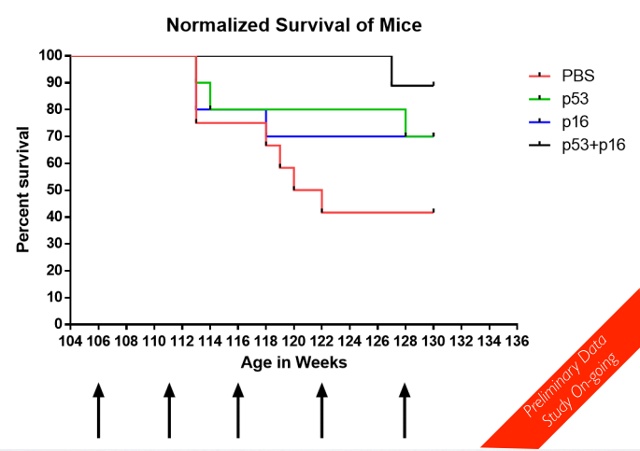
Endothelial Cell Dysfunction as the Early Manifestation of Small Vessel Disease

Cerebral small vessel disease is a form of age-related dysfunction in the smaller blood vessels of the brain, associated with damage to the white matter of the brain and the onset of dementia. It is thought that the increased blood pressure of hypertension and consequent physical stresses on blood vessel walls is the primary cause of small vessel disease, but here researchers provide evidence pointing towards specific forms of change in signaling generated by dysfunctional endothelial cells that form the blood-brain barrier in blood vessel walls. That signaling degrades some of the necessary supporting operations of cells in nearby brain tissue - a situation that sounds similar to the outcome of cellular senescence and the senescence-associated secretory phenotype, though that topic isn't mentioned at all in this paper. This endothelial cell signaling occurs prior to other aspects of small vessel disease, though itself must still be secondary to the underlying molecular damage of aging in and around blood vessel cells: senescence, cross-linking, and so forth.

Cerebral small vessel disease (SVD) affects arterioles in the brain, increasing risk of stroke and causing symptoms of dementia. Magnetic resonance scans of SVD patients typically show white matter abnormalities, but we do not understand the mechanistic pathological link between blood vessels and white matter myelin damage. Hypertension is suggested as the cause of sporadic SVD, but a recent alternative hypothesis invokes dysfunction of the blood-brain barrier as the primary cause.

In a rat model of SVD, we show that endothelial cell (EC) dysfunction is the first change in development of the disease. Dysfunctional ECs secrete heat shock protein 90α, which blocks oligodendroglial differentiation, contributing to impaired myelination: vascular tight junctions of the blood-brain barrier were impaired in SVD, and dysfunctional endothelial cells prevented oligodendrocyte precursors from maturing into myelinating cells.

Treatment with EC-stabilizing drugs reversed these EC and oligodendroglial pathologies in the rat model. EC and oligodendroglial dysfunction were also observed in humans with early, asymptomatic SVD pathology. We identified a loss-of-function mutation in ATPase11B, which caused the EC dysfunction in the rat SVD model, and a single-nucleotide polymorphism in ATPase11B that was associated with white matter abnormalities in humans with SVD. We show that EC dysfunction is a cause of SVD white matter vulnerability and provide a therapeutic strategy to treat and reverse SVD in the rat model, which may also be of relevance to human SVD.

Link: https://doi.org/10.1126/scitranslmed.aam9507

Senolytics Reduce Age-Related Dysfunction and Extend Remaining Life Span by 36% Following Administration to Old Mice

This paper isn't open access, but is important enough to stand out from the many publications on clearance of senescent cells emerging these days. While the evidence is compelling for senescent cells to be a root cause of aging, and removal of senescent cells via senolytic therapies to reverse aspects of aging, many of the fine details remain to be robustly established in the science and the implementations. Measures of senescent cell levels are not yet advanced enough for clinical implementations, for example, and the life span studies have so far involved animals genetically modified to suppress senescence rather than administration of senolytics to normal animals. That has now changed: in this study, researchers demonstrate that the senolytic mix of dasatinib and quercetin produces noteworthy results on the remaining life span of old mice.

Physical dysfunction and incapacity to respond to stresses become increasingly prevalent toward the end of life, with up to 45% of people over the age of 85 being frail. The cellular pathogenesis of age-related physical dysfunction has not been fully elucidated, and there are currently no root cause-directed, mechanism-based interventions for improving physical function in the elderly available for clinical application. Here we report a potential strategy for addressing this need: reducing senescent cell burden.

Senescent cell burden increases in multiple tissues with aging, at sites of pathology in multiple chronic diseases, and after radiation or chemotherapy. Senescent cells can secrete a range of proinflammatory cytokines, chemokines, proteases, and other factors; together, these are termed the senescence-associated secretory phenotype (SASP), which contributes to local and systemic dysfunction with aging and in a number of diseases.

Here we demonstrate that transplanting relatively small numbers of senescent cells into young mice is sufficient to cause persistent physical dysfunction, as well as to spread cellular senescence to host tissues. Transplanting even fewer senescent cells had the same effect in older recipients and was accompanied by reduced survival, indicating the potency of senescent cells in shortening health- and lifespan.

The senolytic cocktail, dasatinib plus quercetin, which causes selective elimination of senescent cells, decreased the number of naturally occurring senescent cells and their secretion of frailty-related proinflammatory cytokines in explants of human adipose tissue. Moreover, intermittent oral administration of senolytics to both senescent cell-transplanted young mice and naturally aged mice alleviated physical dysfunction and increased post-treatment survival by 36% while reducing mortality hazard to 65%. Our study provides proof-of-concept evidence that senescent cells can cause physical dysfunction and decreased survival even in young mice, while senolytics can enhance remaining health- and lifespan in old mice.

Link: https://doi.org/10.1038/s41591-018-0092-9

Blind Upon the Eve of Apotheosis

Our present age of technology and accelerating progress is the first of its kind for our species. It commenced a few short centuries ago with subtle changes in wealth, agriculture, and life expectancy that compounded to form the foundations for the Industrial Revolution. It was a dramatic break with millennia in which stasis or the cyclic advance and retreat of applied knowledge - of civilization - were the norm. In our era, it is instead the case that progress today reliably creates the potential for greater and faster progress tomorrow. The most important consequence of this is arguably not that we now lead rich lives of greater wealth, capacity, and comfort, but that the technologies of tomorrow will radically transform our selves and our nature: these are the last decades of humanity as we know it. Human nature and the human condition as it has existed since the Great Leap Forward, some 50,000 years ago, will become malleable. We will be able to improve upon the human body and human mind. What comes next is something far greater than humanity, both for the billions of individuals who see the transformation from end to end and for our societies as a whole.

It has been argued that we have already changed ourselves greatly through technology. That the humans of Ur were far removed from the small and violently suspicious bands of humans who coexisted with their Neanderthal near relatives. That today's humans of internet and mobile phones are far removed from the humans of Ur and earlier cities. That technologies such as writing and global telecommunications, or even simply the size and density of population, leads to very different minds grown from the same genetic basis. On the face of it, this seems unlikely, however. Upon the eve of apotheosis, in a world linked by networks of communication and rapidly advancing technology, a glance at the activities of humanity finds any given populace worshipping civic idols and chieftains, in ways that are in essence little different from those of the ancient world, or reencting the politics of the greens and the blues of the Byzantine empire. Further, the past century has seen any number of small bands of humans brought into the modern era by neighboring peoples, with no signs of any fundamental difference in their human nature as a result of comparative isolation and lack of technology.

Past technological progress has not changed what it is to be human, the shape of our minds. What comes next will be very, very different in scope and outcome. It will start with some combination of a progressive reverse engineering of the brain, advanced biotechnology capable of altering and improving upon existing organ function, the development of interfaces between neural architecture and computing hardware, and software emulation of functional neural tissue. All of these lines of research are well established today, albeit in comparative infancy. They will converge into the ability to run minds in hardware and software, to alter the way in which minds function, to extend biological minds, and to move them into hardware, piece by piece. We presently forget 98% of everything we experience. That will go away in favor of perfect, controllable, configurable memory. Skills and knowledge will become commodities that can be purchased and installed. We will be able to feel exactly as we wish to feel at any given time. How we perceive the world will be mutable and subject to choice. How we think, the very fundamental basis of the mind, will also be mutable and subject to choice. We will merge with our machines, as Kurzweil puts it. The boundary between mind and computing device, between the individual and his or her tools, will blur.

Over the course of the 21st century, people will have access to an increasing array of options when it comes to enhancing the function of the mind and the body. The young of today will live to see all of that span, and more. Aging will soon become a treatable medical condition, its causes addressed by therapies that repair the molecular damage that accumulates in old tissues. The human genome will become fluid, the subject of any number of treatments that alter genes or gene expression in adult tissues in order to achieve specific benefits such as greater muscle mass or increased resistance to disease. Somewhere down the line, biotechnology and nanotechnology will merge to produce technologies such as artificial cells and cell-like machines, vastly more efficient at specific tasks than their biological counterparts. Biology will become an option, rather than the present mandate. People will be able to move to a more resilient vehicle for the brain than the present human body, and even the brain may be swapped out for better hardware, through a slow process of replacement and integration, one neuron at a time exchanged for a nanomachine.

Piece by piece, we will be able to choose physical immortality. Choose our physical forms. Choose exactly how our minds function. Choose how we think and feel. Choose exactly what being human is for each of us. A great branching in the diversity of minds and appearances will spring forth from our present uniformity in a matter of mere decades. The population may well expand greatly as well; minds in hardware and software can be copied. There are no limitations on the pace of reproduction in such a world, nor would such minds necessarily need to consume anywhere near the same resources as present humans. If we choose to believe that acceleration in technological development is, at root, largely a function of total human population and the degree to which people communicate with one another, then departing from our biological roots will enable the acceleration to continue far past its present limits.

That isn't just a matter of population, however. The pace of progress today bumps up against the limits imposed by organization of efforts, in that it takes a few years for humans to digest new information, talk to one another about it, decide on a course of action, gather together a group, raise funds, and start working. There is no necessary reason for any of these parts of the process to take more than a few seconds, however: consider a world in which human minds run far faster, because they run on something other than biological neurons, because they run hundreds of distinct streams of consciousness simultaneously, and because they are augmented by forms of artificial intelligence that take on some of the cognitive load for task assignment and decision making.

It is, frankly, hard to even speculate about the potential forms taken by society in such an environment. Technology clearly drives human organizational strategies and struggles, for all that the minds of prehistory, of Ur, and of our modern times are all the same. In the past, evolution of society was largely shaped by the ability to communicate over distances and by the size of the population. In the future it will be shaped to a far greater extent by the way in which intelligences think and feel, and the way in which their minds depart from the present standard for human nature. We struggle to model human action in the broadest sense of economic studies, and I suspect that this will be true for any society of minds, no matter how capable they are. The complexity of the group always exceeds the capabilities of any individual or research effort within that group. We can do little more than point out incentives and suggest trends that are likely to emerge from those incentives.

Nonetheless, the transition to what comes after humanity will come to pass, at some pace. Not as rapidly as some would like, in part because organizational matters will continue to happen slowly until minds are enhanced. Yet those of us who are young enough or fortunate enough to see the transition from end to end, perhaps with the aid of the first functional anti-aging therapeutics, will have the opportunity to become entities so capable, different, and vast in comparison to present humans that our ancestors would have called them gods or spirits. It will happen gradually, step by step, each such step forward a sensible choice to participate in an enhancement that brings benefits or a desired change, but in the end the sum of it will indeed be an apotheosis. Humans will become what they desire to become, leaving humanity as we presently understand it far behind. The present human condition is a seed, a childhood, and it will just as inevitably come to an end as we grow to reach our true potential.

The eyes of the world are by no means as closed to this future as was the case even a few short decades ago, when transhumanism was a niche vision. Posthumanity has been explored in fiction, discussion, and research far more extensively. Yet most people live in the here and the now, and act as though next year will be same as this year. It is a strange state of mind given that we are so evidently alive in a time of rapid change. Most of us have passed through the development of personal computing, the internet, and pervasive telecommunications, and have personal points of comparison for the enormous changes to habit and capabilities that have taken place. Equally, most of us squabble over politics, the blues and the greens again, save for retirement, and otherwise in thought and deed anticipate a life that has the same trajectory and span as that of our grandparents. Becoming gods is not on the agenda, not in the plan. It is still inevitable, however. Few will turn down longer lives, perfect memory, immunity to disease, the ability to run multiple streams of consciousness, and much more, when those capabilities exist. Scientists will build the basis for each incremental advance, entrepreneurs will bring it to the masses, and people will choose to better themselves.

Perhaps it will come slowly, perhaps rapidly. But insofar as there is godhood ahead, most will stumble into it without that ever being the intended goal. It is a strange thing to consider, this future of accidental deities, spawned from our largely blind society of people near entirely focused on unrelated minutiae. Does it even make much difference, we might ask, to stand with eyes open and see what is coming?

Cleara Biotech Launches to Develop Senolytic Therapies Based on FOXO4-DRI

The accumulation of senescent cells is thought to be one of the root causes of aging, and a growing body of evidence points to their direct contribution to numerous age-related conditions. Removing senescent cells is a narrow form of rejuvenation, capable of turning back measures of aging and age-related disease. Last year researchers published data on an approach to selective destruction of senescent cells based on interfering in the interaction between FOXO4 and p53. This pushes senescent cells into the form of programmed cell death known as apoptosis, while doing next to nothing to normal cells. The method of interference involves creating a safe but broken version of the FOXO4 protein, FOXO4-DRI, and introducing it into the body. A sizable fraction of senescent cells relying on the presence of working FOXO4 are destroyed as a result. The principal researcher involved in this work has now started a company with the assistance of Apollo Ventures to further develop this line of work into a viable senolytic therapy.

Cleara Biotech has raised seed funding to advance a program that reversed aspects of aging in mice. The modified FOXO4-p53 interfering peptide program made headlines last year when it restored the physical fitness, hair growth, and kidney function of mice. Peter de Keizer, Ph.D., and his collaborators achieved the improvements by targeting cells that had entered senescence, a state in which they stop dividing and start secreting different factors. Studies have linked these cells to an array of big diseases, attracting multiple research groups and powering Unity Biotech to an $85 million IPO. Among all these activities, Keizer's peptide stood out because it eliminated senescent cells without harming healthy tissues.

James Peyer, Ph.D., managing partner at aging-focused fund Apollo Ventures had been looking for a marker specific to senescent cells without success. Such specificity is vital if a drug is to treat chronic, age-related conditions such as kidney disease without causing intolerable side effects. When Peyer's fund saw Keizer's preliminary data, he teamed up with the senescence scientist and his collaborators. The result is Cleara. Cleara will spend the next year optimizing the peptide Keizer tested in mice in anticipation of moving into the clinic. This will entail designing multiple candidates with strengths, pharmacokinetic profiles, and other characteristics tailored to indications that Cleara may target.

Cleara is still zeroing in on indications, with Peyer noting that this is "one of the core challenges for a number of different drugs in this space, where you're presented with a cornucopia of options." But it has a broad idea of the areas it is going to target. One lead optimization strand will develop a candidate against a chronic condition, such as kidney disease, osteoarthritis, or COPD. The second strand will target an acute, life-threatening "rare or rare-ish" disease. This second strand will likely get into the clinic first - reflecting the higher tolerance for risk among patients with life-threatening diseases - and may ultimately target a type of cancer.

Link: https://www.fiercebiotech.com/after-breathing-new-life-into-old-mice-senescence-startup-cleara-bags-cash-to-move-toward-clinic

Chimeric Antigen Receptor Therapy, but Using Natural Killer Cells

Adding chimeric antigen receptors to T cells (CAR-T), causing them to aggressively target cancer cells, has proven to be a fruitful approach to the treatment of cancer. Like most immunotherapies, it can result in potentially severe side-effects related to excessive immune activation, but it is also quite effective. Treatment of forms of leukemia in particular has produced good results in a large fraction of patients who have trialed the therapy. In the research reported here, scientists extend the chimeric antigen receptor approach to natural killer cells rather than T cells, noting that this may prove to be both safer and logistically easier to deploy to large numbers of patients.

Genetically engineered T cells that destroy cancer cells have proven to be promising options when other treatments fail. However, there's currently no one-size-fits-all CAR T, and each patient needs his own bespoke intervention. Now, researchers report that natural killer cells, grown from human induced pluripotent stem (IPS) cells and modified in a similar way to CAR-T cells, are effective against ovarian cancer in a mouse model. The result paves the way for developing an "off the shelf" immunotherapy that doesn't need to be personalized for each patient. Natural killer (NK) cells, which play an important role in tumor surveillance, offer a key advantage over T cells in that they kill some cancer cells without requiring tumor-specific cell-surface receptors, meaning they can work in many patients.

CAR-T cell therapies are built by harvesting a patient's T cells and genetically modifying them to produce so-called chimeric antigen receptors (CARs) that direct them to destroy cancer cells. Two such immunotherapies were approved last year. Unmodified NK cells isolated from peripheral blood or umbilical cord blood have also been shown to be effective against acute myelogenous leukemia in several clinical trials, and a few trials testing NK cells equipped with CARs in other forms of blood cancer have begun. But developing a means of deriving NK cells from stem cells would allow researchers to generate hundreds of thousands of doses that are standardized.

The team created the mouse models by transplanting human ovarian cancer cells into mice whose immune systems had been suppressed to prevent them from rejecting the human cells. The scientists then infused the CAR-NK cells into the animals, and for comparison, did the same for CAR-T cells. They noted that the mice treated with the iPSC-derived CAR-NK cells and those treated with CAR-T cells both had shrunken tumors after 21 days. The researchers were surprised to find that, compared to the mice treated with CAR NKs, the animals that had received the CAR-T cell treatment appeared to be in worse shape: they had damage in organs such as the liver, lungs, and kidneys and an increase in inflammatory cytokines. "The mice that got the CAR-T cells actually wound up getting sick, losing weight, and getting these toxicities, whereas the CAR-NK-cell-treated mice didn't."

Link: https://www.the-scientist.com/news-opinion/natural-killer-cells-prove-effective-as-a-car-therapy-in-mice--64456

The Humble Axolotl and the Quest for Human Organ Regeneration

The popular science article I'll point out today takes a look at research into axolotl biochemistry. The scientists involved are searching for ways in which they might be able to improve upon mammalian regeneration; the axolotl is one of the few higher species capable of perfect, repeated regeneration of lost limbs and severe damage to other organs. There are limits, of course, and the axolotl is just as mortal as any mammal, but mammals, ourselves included, have in comparison a very poor capacity for regeneration. We can barely grow back a fingertip, and even that only when very young, and not at all reliably. There are tantalizing hints that the capacity for far greater feats of regeneration still lurks within mammals, but disabled, or overridden. Mammals can regenerate during later embryonic development. The MRL mouse lineage can regenerate small injuries without scarring. African spiny mice have evolved to regenerate whole sections of skin perfectly, and don't appear all that different from other rodents in other aspects.

In addition to axolotls, researchers work with zebrafish, newts, and other highly regenerative species. It is an exercise in comparative biology, an effort to reverse engineer the important differences between these species and mammals. Some interesting advances have emerged in recent years. For example the human tumor suppressor ARF shuts down the exceptional regeneration of zebrafish when introduced into that species. This strongly suggests that the majority of higher species that have poor regenerative capacity do so because of evolved defenses against cancer. After all, the controlled cell growth of regeneration versus the uncontrolled cell growth of cancer are two sides of the same coin. Another important discovery centers around the role of macrophages and transient senescent cells in regeneration. The details of the intricate interactions between these and other cell types in injured tissue seems at the center of the choice between scarring or regrowth. Mammals scar, axolotls regrow.

Despite being a focus of attention for the popular science media, the prize here is not really a way to regrow lost limbs. That is a minor benefit. The real prize is a way to repair damage to internal organs, including important aspects of the slow-moving loss of function that arises with age, such as the internal scarring of fibrosis. Enabling axolotl-like proficient regeneration will be a form of therapy that partners well with the restoration of stem cell function in the old. Both lines of research may come to fruition in the clinic on a similar timescale in the years ahead.

Salamander's Genome Guards Secrets of Limb Regrowth

In a loudly bubbling laboratory, about 2,800 of the salamanders called axolotls drift in tanks and cups, filling floor-to-ceiling shelves. Salamanders are champions at regenerating lost body parts. A flatworm called a planarian can grow back its entire body from a speck of tissue, but it is a very small, simple creature. Zebrafish can regrow their tails throughout their lives. Humans, along with other mammals, can regenerate lost limb buds as embryos. As young children, we can regrow our fingertips; mice can still do this as adults. But salamanders stand out as the only vertebrates that can replace complex body parts that are lost at any age, which is why researchers seeking answers about regeneration have so often turned to them.

While researchers studying animals like mice and flies progressed into the genomic age, however, those working on axolotls were left behind. One obstacle was that axolotls live longer and mature more slowly than most lab animals, which makes them cumbersome subjects for genetics experiments. Worse, the axolotl's enormous and repetitive genome stubbornly resisted sequencing. Then a European research team overcame the hurdles and finally published a full genetic sequence for the laboratory axolotl earlier this year. That accomplishment could change everything.

After an amputation, a salamander bleeds very little and seals off the wound within hours. Cells then migrate to the wound site and form a blob called a blastema. Most of these recruits seem to be cells from nearby that have turned back their own internal clocks to an unspecialized or "dedifferentiated" state more like that seen in embryos. But it's unclear whether and to what extent the animal also calls on reserves of stem cells, the class of undifferentiated cells that organisms maintain to help with healing. Whatever their origin, the blastema cells redifferentiate into new bone, muscle and other tissues. A perfect new limb forms in miniature, then enlarges to the exact right size for its owner.

Arms, legs, and tails aren't the only body parts that laboratory axolotls can regrow. They also recover from crushing injuries to their spinal cords. They can regenerate a millimeter-by-2-millimeter square of their forebrain. Scientists don't know whether axolotls use the same mechanisms to regenerate their internal organs as their limbs. They also don't know why an axolotl can grow back an arm many times in a row but not indefinitely - after being amputated five times, most axolotl limbs stop coming back. Another mystery is how a limb knows to stop growing when it reaches the right size. But these may not be mysteries for much longer.

Biomedical Engineering in Medicine and Aging

Today, the effective treatment of aging can only proceed rapidly as an engineering project. The fine details of the way in which aging progresses at the level of cells and proteins are far from fully understood - but that is not a roadblock to progress. The research community knows enough of the causes of aging to repair them and observe the results. In fact the repair approach, where it has been tried, and as typified by senolytic development to clear senescent cells, is doing far more, with far less expenditure, and in far less time, than other strategies that involve mapping and adjusting the extreme complexity of cellular metabolism.

A good analogy for this situation is that sizable bridges and other large structures were constructed on an empirical basis for millennia prior to a full understanding of materials science, prior to the implementation of computational modeling in architecture. In exactly the same way it is possible to make meaningful progress in the treatment of aging today, and because aging causes far more harm than any other aspect of the human condition, it is our ethical imperative to make that progress rather than waiting on greater understanding of how exactly aging and metabolism interact in detail.

Biomedical gerontologist Aubrey de Grey says he believes that, thanks to imminent advances in technology, the first person to live to age 1,000 likely is alive today. De Grey became interested in studying the biological aspects of aging, he says, because he views its negative effects, such as chronic pain and memory loss, as preventable. While he is focused on improving quality of life, he says longevity is a "side effect" of good health. He helped to found the SENS (Strategies for Engineered Negligible Senescence) Research Foundation, a regenerative medical research nonprofit that focuses on preventing diseases and disabilities related to aging.

Living much longer lives will happen soon, de Grey contends, challenging the notion that aging is inevitable or even natural. Radiologist Joon Yun agrees. Yun is president and managing partner of Palo Alto Investors. His family foundation has sponsored a number of initiatives and made a US $2 million gift to launch the National Academy of Medicine's Healthy Longevity Grand Challenge, to "solve aging" as he puts it. Like de Grey, he says he is focused on improving quality of life so that people will be able to continue their active lifestyles without any trouble, no matter their age.

There are many opportunities in tissue engineering, stem-cell therapy, and immunotherapy. One of the projects the SENS Research Foundation is working on involves "death resistant cells," which cause degenerative aging. These cells cause loss of muscle mass, inflammation, and metabolic changes. The foundation is looking into rejuvenating cellular and molecular structures to keep people young. To restore health and vigor, organizations partnering with the foundation are experimenting with growing organs and cells in labs. That could allow for organs to be custom-made from the recipient's cells. The cells also could be used as neurons for the brain or muscle for the heart. The process might eliminate the need for organ donors or searching for a match. It also would reduce the risk of transplant rejection, and a recipient's organs would be biologically "younger" than those from a donor.

As people get older, they experience a decline in such vital functions as memory, digestion, and blood circulation. Yun is focused on what he calls functional longevity. He is offering a $1 million cash prize to researchers who can "hack the aging code," or find a solution to aging that would prolong lives and maintain quality of life. Yun's approach to prolonging life span is to restore the body's ability to respond to stress, known as homeostatic capacity. As we get older, our capacity declines, causing functions to weaken. Yun is looking for entrepreneurs and engineers who have ideas on how to measure homeostatic capacity. "Rather than thinking about inflammation as a cause of aging, we should think about it as a loss of inflammatory capacity. Instead of thinking that weight gain is just part of aging, think of it as a loss of metabolic capacity."

Link: http://theinstitute.ieee.org/career-and-education/career-guidance/biomedical-engineers-look-to-help-people-live-much-longer

Reducing Levels of All Pathological Protein Aggregates Should be a Primary Strategy for Treating Neurodegeneration

The reasons why restoration of cerebrospinal fluid drainage is a very promising strategy for the treatment of Alzheimer's disease go beyond the compelling direct evidence, into matters of research and development strategy. Numerous proteins that become misfolded or altered in ways that cause them to form solid deposits in the aging brain, surrounded by a halo of harmful secondary biochemistry. To date, serious development efforts that have advanced to clinical trials have focused on clearing only one of these aggregates. That may well never be enough: neurodegeneration appears to be a combination of the effects of many mechanisms of similar weight and consequence.

Thus more researchers are beginning to call for broader efforts that target multiple problem proteins. In this context, the importance of improved cerebrospinal fluid drainage is that it can can reduce the levels of all molecular waste and resulting consequences in the brain, both the well understand and the less well understood alike. It all flows out through the same channels, provided that those channels are working well enough. Unfortunately they decline with age, and that is a comparatively simple, mechanical and structural potential point of intervention. I am looking forward to the data produced by Leucadia Therapeutics as their metholodology for drainage restoration progresses over the years ahead: they should be able to fairly quickly definitively confirm the utility of this strategy.

Nearly all major neurodegenerative diseases - from Alzheimer's to Parkinson's - are defined and diagnosed by the presence of one of four proteins that have gone rogue: tau, amyloid-beta (Aβ), alpha-synuclein (α-syn), or TDP-43. As such, investigational drugs and studies aimed at preventing or slowing the disease often hone in on just one of these respective proteins. However, targeting multiple proteins at once may be the real key, according to a recent study. These so-called "proteinopathies" - misfolded proteins that accumulate and destroy neurons - co-exist in varying degrees across all of the different neurodegenerative disorders and may instigate each other to drive disease severity in many aging patients. The prevalence of these co-pathologies suggests that each disease may ultimately require combination therapy targeting multiple disease proteins, and not just a single therapy, in patients with both early and later-stage disease.

"Historically, the focus of most clinical trials has been on targeting the primary pathological proteins of a given neurodegenerative disease such as deposits of tau and Aβ for Alzheimer's disease, but we see now that many of these disease-related aggregated proteins affect most older patients across a full spectrum of clinical and neuropathological presentations. This gives us additional leverage to find ways to detect patients' specific proteinopathies with increasingly sophisticated biomarker and imaging technologies. This will allow us, and other researchers, to better match participants with specific targeted therapies in clinical trials."

The study analyzed 766 autopsied brains and revealed that patients with more severe forms of their diseases had more co-pathologies. Co-pathologies were common but varied among the disease groups, ranging from 27 to 81 percent of patients having co-pathologies. For example, 52 percent of patients with corticobasal degeneration (CBD), in which tau as the primary protein, had multiple other neurodegenerative disease protein deposits present. Tau was nearly universal, with 92 to 100 percent of all patients having at least one form. Aβ was next, with 20 to 57 percent of patients having at least one type of protein deposit, while α-syn pathology, typically seen in Parkinson's disease, was less common, with 4 to 16 percent. TDP-43 deposits, which are characteristic pathological signatures of frontotemporal lobar degeneration and amyotrophic lateral sclerosis, were the rarest, with 0 to 16 percent of patients having these deposits.

The findings not only show a high prevalence of co-pathologies, but also suggest a patient's primary pathological protein may influence co-pathology prevalence and severity, as shown in patients with Alzheimer's and Lewy body disease patients. These findings support the "proteopathic seeding" hypothesis that has been previously established in model systems of neurodegenerative diseases. Misfolded proteins may directly "cross-seed" other normal, vulnerable proteins to accumulate and clump via a cell-to-cell transfer of toxic proteins.

Link: https://www.pennmedicine.org/news/news-releases/2018/july/rethinking-neurodegenerative-disease-treatment-target-multiple-pathological-proteins

Can the Top-Down Institutional Approach Promote the Right Sort of Research and Development to Treat Aging?

Most scientists who spend their professional lives within large institutions, such as the big universities, the National Institute on Aging (NIA), and so forth, tend to favor institutional solutions. In practice that means slow engineering of change within the established hierarchy, rather than stepping outside it, or where a new need is identified, meeting it with the creation of a new institutional edifice much like those that already exist. This is the top down approach to development: structure and delegation, provide big-picture guidance and leave the details up to lower levels of the hierarchy. It is advocated in a recent open access position paper authored by a noted Russian researcher, one of those who led the development of plastinquinone based mitochondrially targeted antioxidants as a potential means to slow the progression of aging.

It is necessary to establish an International Agency for Research on Aging

Extending healthy lifespan is one of the main goals of gerontology and preventive medicine. There are potential interventions which might delay and/or prevent the onsets of many chronic pathologies associated with human aging. The affected pathways have been identified, and the behavioral, dietary, and pharmacologic approaches to preventing and treating age-related disorders have emerged. Interventions that target the aging process in its entirety appear to be more effective in preventing a broad range of age-related pathologies than specific interventions targeting such pathologies. Development of the new anti-aging drugs opens broad prospects for the pharmaceutical and healthcare industries. However, if human longevity continues to advance, the incidences of age-associated diseases, including cardiovascular diseases, type 2 diabetes, and cancer would also increase thus presenting a tremendous challenge for humankind. The search for adequate models for selecting the effective and safe methods of healthy life extension has become a priority in biology of aging.

There are at least two broadly accepted definitions of pharmacological compounds capable of intervening in aging: a) anti-aging drugs, which presumably reverse the aging process ('rejuvenation'), and b) geroprotectors, which are supposed to prevent premature aging and/or slow down or postpone aging. The term "longevity therapeutics" has been introduced for drugs that can interfere with the process of aging and extend the mean and/or maximum lifespan, preserve physiological functions and mitigate the onset and severity of a broad spectrum of age-associated diseases in mammals. Potential geroprotective agents have been subdivided into several groups: those that demonstrate an anti-aging effect without any evidence of lifespan increase; those that increase lifespan by reducing the incidence of age-associated pathology; and those that extend lifespan presumably by reversing the aging process itself. Most of the evidence related to these definitions and classifications has been gained in animal studies.

The designs of most such studies were found to have various deficiencies which led to confounding results. Therefore, there is the need to work out standard guidelines for testing such drugs and evaluating their life extending potential as well as various late effects, including tumor development. Guidelines for testing should include such significant elements as animal models, testing regimens, and biomarkers/endpoints. To this end, it is necessary to develop international standards for conducting the preclinical and clinical studies of agents intended to be used in pharmacological interventions in aging, as well as for evaluating the results of such studies. In the years to come, a promising agenda could be the development of new biomarkers based mostly on biochemical and genetic tests for short-term screening of potential agents. Collaborative studies of anti-aging drugs and geroprotectors conducted in various laboratories could be particularly promising.

In 2000, an international program on the assessment of the efficacy and safety of geroprotectors was proposed. It was suggested that the proposed program could be established under the auspices of the United Nations Program on Aging, the World Health Organization, and the International Association of Gerontology and Geriatrics. Unfortunately, the proposed program has not been implemented. We believe that it is worth reverting to that earlier proposal. Whereas the main challenge for healthcare in the 20th century had been the rapid increase in morbidity and mortality from malignant neoplasms, in the 21st century the primary challenge will be the effects of global aging on public and individual health. Therefore, the establishment of an International Agency for Research on Aging (IARA) under the auspices of the World Health Organization, similar to the International Agency for Research on Cancer (IARC), is expedient. Similarly to IARC, the objective of IARA should be the promotion of international collaboration in gerontology and geriatrics.

Can this really work, however? The challenge with large institutions is that the natural human inclination to conservatism, to playing it safe, to avoiding change, is magnified tenfold. If you want to maintain the status quo, that might be great, but if you want to change the world, then institutions are usually the enemy. The NIA mission involves "understanding the nature of aging and the aging process, and diseases and conditions associated with growing older, in order to extend the healthy, active years of life." This organization has been in existence since the 1970s; why are we still aging at much the same pace, with none of the fundamental causes meaningfully addressed? All of this funding has certainly led to the accomplishment of a great deal of scientific work, but sadly next to nothing of practical use when it comes to ways to slow or reverse the aging process. This isn't because they have no starting point: many of the root causes of aging have been well described for decades. Yet there is absolutely no danger that the NIA will meaningfully support the best directions for rejuvenation in the near future.

Much the same is true of other large institutions. They favor examination of aging, small changes to aging achieved by tinkering with metabolism, or the safe old school path of palliative methods of making age-related disease slightly less worse. Few exhibit any interest in the well known potential approaches to reversing aging by repairing its root causes. The World Health Organization won't even acknowledge that aging can be treated at all! What one can expect from a sizable institution charged with a specific mission is for its functionaries to pick the smallest possible set of changes they can aim for, and then work ineffectually to achieve those changes.

Senolytic rejuvenation therapies are being pioneered outside the institutions, as they did not support it despite the decades of evidence. The Methuselah Foundation and SENS Research Foundation, instrumental in steering the research community towards better strategies, were created by outsiders because the institutions of aging research would not acknowledge the need and the opportunity for effective rejuvenation development programs. Revolution and new, useful paths forward near always arise outside the mainstream, and are opposed by establishment institutions for as long as possible. That certainly happened, and is still in the process of happening, for rejuvenation research. So I'd say that the support we give to a better future is best directed to bottom-up approaches. Rebels, revolutionaries, startups, and rejuvenation biotechnology. New ideas, new directions, not the same old careful preservation of the status quo that exists in the largest research bodies.

Differences in Macrophage Polarization Between More Healthy and Less Healthy Elders

Macrophages of the innate immune system take on different states known as polarizations depending on their duties. M1 macrophages are aggressive and inflammatory, involved in the destruction of pathogens and harmful cells. M2 macrophages aid in the processes of regeneration. The immune system becomes more inflammatory with advancing age. This chronic inflammation drives progression of most of the common age-related diseases, and an excess of M1 macrophages appears as a feature of many of those conditions. The research community is looking into ways to force more macrophages into the M2 polarization, as a possible approach to override a fraction of age-related immune dysfunction. In this context, researchers here report on their evaluation of differences in the macrophage population between more and less healthy older individuals.

The low-grade, chronic inflammatory state affecting aging organisms - inflammaging - is among the major risk factors for the development of the most common human age-related diseases (ARDs). Increasing evidence suggests that inflammaging is underpinned by monocytes/macrophages, while the acquisition of a senescent phenotype - a phenomenon that has been defined as macroph-aging - impairs the ability of immune cells to cope with stressors, thus contributing to immunosenescence. In this framework, the role of macrophage polarization in the modulation of inflammatory and repair processes is attracting growing interest.

The two main, and opposite activities of macrophages have led them to be classified into pro-inflammatory classically activated macrophages (M1) and anti-inflammatory and immunoregulatory alternatively activated macrophages (M2). Despite its value, this classification is however insufficient to describe the diverse phenotypes and functions of monocytes/macrophages in vivo. Intense research is being devoted to associate the polarization profiles, seen in vitro in relation to specific stimuli, with circulating and/or tissue macrophage polarization in health and disease conditions. Clearly, in vitro models are unable to mimic the complex environment that influences the M1/M2 balance in vivo, and since macrophages can develop mixed M1/M2 phenotypes, novel in vivo detection strategies are required. Several biomarkers have been associated with M1/M2 profiles. CD163, the high-affinity scavenger receptor for the haemoglobin-haptoglobin complex, is selectively expressed on M2 macrophages and monocytes, whereas CD80, a costimulatory signal for T cell activation and survival, is preferentially expressed on M1 macrophages.

Since data on the M1/M2 phenotype of circulating monocytes in healthy aging are not available, this study was undertaken to analyse monocyte profiles in healthy subjects of different ages using flow cytometry. To establish whether an M1/M2 imbalance could be disease-associated, the M1/M2 phenotype of elderly healthy subjects was compared with the one of elderly patients with acute myocardial infarction (AMI). The AMI patients showed a significantly decreased proportion of CD163+CD80+ and an increased proportion of CD163+ and CD163-CD80- cells among classical monocytes, opposite trends to those observed in healthy aging. Moreover, a significantly greater proportion of intermediate and non-classical CD80+ monocytes suggested a shift to a pro-inflammatory phenotype. Overall, CD163/CD80 characterization of circulating monocytes provides additional information about their polarization and could be an innovative tool to monitor aging.

Link: https://doi.org/10.18632/aging.101465

Metformin Shown to Attenuate Lung Fibrosis in Mice

Fibrosis is a form of malfunction in tissue maintenance and regeneration, in which cells inappropriately build scar-like collagen structures that disrupt normal tissue function. It is perhaps most significant in age-related diseases of the lung, heart, and kidney, but it is a general feature of old tissues. There are no effective and approved treatments capable of reversing fibrosis to any significant degree, but good evidence has arrived in recent years to suggest that senescent cells, one of the root causes of aging, are also an important contributing cause of the regenerative dysfunction that leads to fibrosis. Senolytic therapies capable of selectively removing senescent cells from an organ with fibrosis should prove helpful.

In that context, it is interesting to look over this recent demonstration of attenuated lung fibrosis via metformin treatment. Metformin is thought to modestly slow aging, being a form of calorie restriction mimetic, but as such treatments go, it is notably poor and unreliable. The animal data is highly varied when it comes to practical outcomes on aging and longevity. The beneficial effect on fibrosis observed here is thought to be mediated via mitochondrial function. Given what is known of metformin in aging, cellular senescence in fibrosis, and the role of mitochondria in programmed cell death, removing problem cells from tissue, it is tempting to speculate on the destruction of a fraction of the senescent cells in a fibrotic organ. But again, we know that metformin is unreliable in animal studies, while senolytics are exactly the opposite. So other mechanisms seem more likely, such as a change in cell behavior prompted by better function in mitochondria.

Pulmonary fibrosis can develop after lung injuries like infections, radiation, or chemotherapy, or it can have an unknown cause, as in idiopathic pulmonary fibrosis, or IPF. IPF is a progressive, and ultimately fatal, lung disorder. In experiments using lung tissues from patients with IPF, mouse lung fibroblasts, and a murine model of lung fibrosis, a team showed the reversal of lung fibrosis and the underlying cellular mechanisms affected via drug treatment. Interestingly, the drug that accelerated the resolution of lung fibrosis is metformin, which is a safe and widely used agent for non-insulin-dependent diabetes.

The research focused on AMP-activated protein kinase (AMPK), an enzyme that senses energy state in the cell and regulates metabolism. Researchers found that AMPK activity was lower in myofibroblast cells within fibrotic regions of human lung tissue from IPF patients. Myofibroblasts deposit extracellular collagen fiber as part of the fibrosis process. These myofibroblasts were metabolically active and were resistant to the programmed cell death called apoptosis, a natural process that removes more than 50 billion damaged or aged cells in adults each day.

Activation of AMPK in myofibroblasts from lungs of humans with IPF, using the drug metformin or another activator called AICAR, led to lower fibrotic activity. AMPK activation also enhanced the production of new mitochondria, the organelles in cells that produce energy, in the myofibroblasts, and it normalized the cells' sensitivity to apoptosis. Using a mouse model for lung fibrosis elicited by the anti-cancer drug bleomycin, the research team found metformin treatment, starting three weeks after lung injury and continuing for five weeks, accelerated the resolution of well-established fibrosis. Such resolution was not apparent in AMPK-knockout mice, showing that the effect of metformin was AMPK-dependent.

"Together, our studies support the concept that AMPK may function as a critical metabolic switch in promoting resolution of established fibrosis by shifting the balance from anabolic to catabolic metabolism. Additionally, we provide proof-of-concept that activation of AMPK by metformin or other pharmacologic agents that activate these pro-resolution pathways may be a useful therapeutic strategy for progressive fibrotic disorders."

Link: http://www.uab.edu/news/research/item/9567-metformin-reverses-established-lung-fibrosis

Prevention of Harmful Astrocyte Activation as a Therapy for Parkinson's Disease

Researchers have recently investigated means to interfere in on a one of the later consequences in neurodegenerative conditions, in which the supporting astrocyte cells in the brain become actively harmful to the neurons that they normally aid and protect. Astrocytes are triggered into this state at least in part by the inflammatory dysregulation of microglia, a class of innate immune cells of the central nervous system. Aging brings rising levels of chronic inflammation throughout the body, a consequence of processes such as the accumulation of senescent cells and malfunctioning of the immune system. The evidence clearly shows that this inflammation contributes to the progression of all of the common age-related conditions, and neurodegenerative diseases are no exception.

The focus in the research materials noted here is on Parkinson's disease, but the mechanism is more broadly applicable. All older individuals suffer to some degree from inflammation of the central nervous system, and the more of it there is, the worse off they are. Sabotaging one of the numerous consequences of this inflammatory state is better than nothing, but it isn't as good as finding ways to address the roots of the issue. In this case, that would be the causes of what has come to be known as inflammaging, the decline of the immune system into simultaneous incompetence and excess activity.

There are a number of strategies that could be pursued effectively today, even given the present poor state of knowledge of the precise cellular and biochemical details of later stage aging. Regeneration of the thymus and replacement of hematopoietic stem cells to restore a youthful supply of immune cells; clearance of the existing immune system to remove malfunctioning and maladapted cells; clearance of senescent cells to remove their inflammatory signals; and so forth. While some companies are working in these areas, all in all too little effort is being directed towards these and related strategies that are in principle capable of turning back immune aging.

Experimental Drug Stops Parkinson's Disease Progression in Mice

NLY01 works by binding to glucagon-like peptide-1 receptors on the surface of certain cells. Similar drugs are used widely in the treatment of type 2 diabetes to increase insulin levels in the blood. Though past studies in animals suggested the neuroprotective potential of this class of drugs, researchers had not shown directly how it operated in the brain. To find out, they tested NLY01 on three major cell types in the human brain: astrocytes, microglia, and neurons. They found that microglia, a brain cell type that sends signals throughout the central nervous system in response to infection or injury, had the most sites for NLY01 to bind to - two times higher than the other cell types, and 10 times higher in humans with Parkinson's disease compared to humans without the disease.

Microglia secrete chemical signals that convert astrocytes - the star shaped cells that help neurons communicate with their neighbors - into aggressive "activated" astrocytes, which eat away at the connections between cells in the brain, causing neurons to die off. Researchers speculated that NLY01 might stop this conversion. In a preliminary experiment in laboratory-grown human brain cells, the researchers treated human microglia with NLY01 and found that they were able to turn the activating signals off. When healthy astrocytes were combined with the treated microglia, they did not convert into destructive activated astrocytes and remained healthy neuroprotective cells.

Researchers tested the drug's effectiveness in mice engineered to have a rodent version of Parkinson's disease. They injected the mice with alpha-synuclein, the protein known to be the primary driver of Parkinson's disease, and treated the mice with NLY01. Similar but untreated mice injected with alpha-synuclein showed pronounced motor impairment over the course of six months in behavioral tests. However, the researchers found that the mice treated with NLY01 maintained normal physical function and had no loss of dopamine neurons, indicating that the drug protected against the development of Parkinson's disease.

Block of A1 astrocyte conversion by microglia is neuroprotective in models of Parkinson's disease

Activation of microglia by classical inflammatory mediators can convert astrocytes into a neurotoxic A1 phenotype in a variety of neurological diseases. Development of agents that could inhibit the formation of A1 reactive astrocytes could be used to treat these diseases for which there are no disease-modifying therapies. Glucagon-like peptide-1 receptor (GLP1R) agonists have been indicated as potential neuroprotective agents for neurologic disorders such as Alzheimer's disease and Parkinson's disease.

The mechanisms by which GLP1R agonists are neuroprotective are not known. Here we show that a potent, brain-penetrant long-acting GLP1R agonist, NLY01, protects against the loss of dopaminergic neurons and behavioral deficits in the α-synuclein preformed fibril (α-syn PFF) mouse model of sporadic Parkinson's disease. NLY01 also prolongs the life and reduces the behavioral deficits and neuropathological abnormalities in the human A53T α-synuclein (hA53T) transgenic mouse model of α-synucleinopathy-induced neurodegeneration.

We found that NLY01 is a potent GLP1R agonist with favorable properties that is neuroprotective through the direct prevention of microglial-mediated conversion of astrocytes to an A1 neurotoxic phenotype. NLY01 should be evaluated in the treatment of Parkinson's disease and related neurologic disorders characterized by microglial activation.

The Longevity Film Competition, Advocacy for Rejuvenation Research

The volunteers at Heales and the International Longevity Alliance last organized a film competition in support of longevity science and longer, healthier lives back in 2015. Now the second competition in this series is underway, with its own website, and the aim of spreading the word about the potential inherent in the treatment of aging as a medical condition. Given enthusiasm and funding, aging might be controlled and halted in the decades ahead, through the development of therapies that repair its root causes. I hope to see a brace of interesting entries as this contest progresses, given the growth in our community over the past few years. If you have an idea and the time to act on it, why not give it a try?

This is an international film competition created to help convey the importance of addressing age related disease. Our jury is composed of filmmakers, scientists, successful entrepreneurs and experts in the fields of regenerative medicine, aging, and longevity. We are living in very interesting times, times of constant change. The scientific community is telling us that soon we could enjoy much healthier and longer lives thanks to technological advancements happening at an accelerated rate. The future can be bright and healthy and we want more people to know about this amazing prospect and want them to get involved in this important mission; the mission of healthy longevity.

However, describing something potentially beautiful is not always easy. We think you can help by making a (very) short movie conveying that a longer and healthier life thanks to sustainable medical interventions, will be a very positive thing for citizens and society alike. Help us spread the word in the right way, help us make sure people understand this is about health and that for the first time in history the possibility of tackling aging is not science fiction, but science fact. Join us in this crusade by entering our competition presented by the SENS Research Foundation, the Healthy Life Extension Society and the International Longevity Alliance and not only potentially help saving lots of lives, but also win the first prize of $10,000! We look forward to your ideas on how to better communicate this important message to the world.

Link: https://longevityfilmcompetition.com/

Adjusting Macrophage Polarization as a Basis for Cancer Immunotherapy

Macrophage polarization is a hot topic of late. The innate immune cells known as macrophages are responsible for a wide range of duties that include destroying errant cells, attacking pathogens, cleaning up waste and debris, and participation in tissue regrowth and regeneration. The polarization of a macrophage describes its state and inclination as to which of those duties it undertakes: M1 macrophages are aggressive and inflammatory, while M2 macrophages tend towards participation in the gentler processes of rebuilding and regeneration. Many of the common inflammatory age-related conditions appear to be characterized by too many M1 macrophages and too few M2 macrophages. Methods by which that balance can be shifted seem promising as a basis for therapy.

Cancer is a different story, however, as is often the case. Many of the issues seen in aging can actually be helpful when it comes to the short-term goals of shutting down and eliminating cancerous tissue. Forcing cancerous cells into senescence is a viable strategy, for example, even though researchers know that senescent cells are one of the root causes of aging, and are working on ways to remove them from normal aged tissues. In this case, the problem of too many M1 macrophages is actually a desirable goal when it comes to attacking cancer. The researchers here are clearly achieving good results via encouraging more macrophages to take up the M1 phenotype in the cancer environment.

Much cancer immunotherapy research has focused on harnessing the immune system's T cells to fight tumors, "but we knew that other types of immune cells could be important in fighting cancer too." Now researchers report that in preclinical models they can amplify macrophage immune responses against cancer using a self-assembling supramolecule. As immune cells, macrophages usually eat foreign invaders including pathogens, bacteria, and even cancer cells, but one of the two types do not always do so. Macrophage type M1s are anti-tumorigenic, but M2s can be recruited by tumor cells to help them grow. Also, tumor cells overexpress a protein that tells the macrophages, "don't eat me." In this way, pro-tumorigenic macrophages may make up 30 to 50 percent of a tumor's mass.

"With our technique, we're re-programming the M2s into M1s by inhibiting the M2 signaling pathway. We realized that if we can re-educate the macrophages and inhibit the 'don't eat me' protein, we could tip the balance between the M1s and M2s, increasing the ratio of M1s inside the tumor and inhibiting tumor growth." The researchers used a multi-component supramolecular system that self-organizes at the nanoscale to deliver an antibody inhibitor plus a drug inside the tumor. This is the first time anyone has combined a drug that targets M2 macrophages and an antibody that inhibits the 'don't eat me signal' in one delivery system.

The researchers tested the supramolecular therapeutic in animal models of two forms of cancer, comparing it directly with a drug currently available in the clinic. Mice that were untreated formed large tumors by Day 10. Mice treated with currently available therapies showed decreased tumor growth. But mice treated with the new supramolecular therapy had complete inhibition of tumor growth. The team also reported an increase in survival and a significant reduction in metastasis. The next steps are to continue testing the new therapy in preclinical models to evaluate safety, efficacy and dosage.

Link: http://www.umass.edu/newsoffice/article/new-cancer-immunotherapy-shows-promise

Cardiac Muscle Cell Therapy Improves Function after Heart Attack in Monkeys

Cell therapies are thought to have great promise as a way to help repair damaged tissue that will not normally regenerate to any great degree. When it comes to the heart, and following nearly two decades of stem cell and other therapies tested in trials and via medical tourism, the research community is still in search of a reliable, highly effective methodology. Work in the laboratory continues, and researchers have recently reported improvement in heart function following heart attack in Southern pig-tailed macaques.

The approach used here involves generating a sizable cell population of cardiomyocytes, heart muscle cells, from embryonic stem cells. Those cells are then introduced into the heart directly, where a large enough proportion of them survive to produce long term reconstruction and some gain in function - not yet a path back to normal, but better than the alternative. The survival of the transplanted cells is the key to effective regenerative therapies: approaches using patches of lab-grown heart tissue in which there is sizable survival of cells following transplantation have also shown promise in heart repair. First generation stem cell therapies are less effective and reliable when it comes to tissue regrowth precisely because near all the cells die, and the benefits produced are mediated by signaling effects on native cells.

The heart is an organ in which the fine structure matters. Its muscle cells participate in an electrical communication network, ensuring that all beat in unison. If that network is disrupted by haphazard growth, then failure of function can result. Unfortunately, there are the first signs of that in this study. The challenge for heart regeneration is perhaps less that it is naturally one of the least regenerative organs, and more that regeneration must be achieved carefully. Other organs are less of a problem on this front, particularly those that are essentially chemical factories or cell nurseries, and their structure and even location isn't anywhere near as important as is the case for the heart or the brain.

Cardiac Cell Transplants Help Monkeys' Hearts

Injecting human cardiac muscle cells into monkeys that suffered heart attacks helped the animals' damaged hearts pump blood better, researchers report. The treatment is based on the reprogramming of human embryonic stem cells, and the results move the therapy a step closer to clinical trials. "We're talking about the number one cause of death in the world for humans. And at the moment all of our treatments are dancing around the root problem, which is that you don't have enough muscle cells."

When a heart attack goes untreated, blood is blocked from flowing to the heart, which leads to the death of heart muscle cells. There can also be scarring and heart failure - when the heart cannot pump enough blood to the body. In the study, after 9 monkeys were made to have heart attacks, their heart-pumping capacity dropped by more than 30 percent. Injecting 750 million cardiac muscle cells, derived from human embryonic stem cells, into the monkeys' hearts led to the growth of new heart muscle tissue. After four weeks, most monkeys' hearts showed improved pumping capacity, up to a third better than right after the heart attacks, and two monkeys had two-thirds of the lost capacity restored after 12 weeks.

However, some of the monkeys had irregular heartbeats after the cardiac cell transfusion. "That is a very important observation because now you can perhaps begin to design a strategy to get at what is happening. How can we prevent this from happening? That, to me, is the story of this paper."

Human embryonic stem cell-derived cardiomyocytes restore function in infarcted hearts of non-human primates

Pluripotent stem cell-derived cardiomyocyte grafts can remuscularize substantial amounts of infarcted myocardium and beat in synchrony with the heart, but in some settings cause ventricular arrhythmias. It is unknown whether human cardiomyocytes can restore cardiac function in a physiologically relevant large animal model. Here we show that transplantation of ∼750 million cryopreserved human embryonic stem cell-derived cardiomyocytes (hESC-CMs) enhances cardiac function in macaque monkeys with large myocardial infarctions.

One month after hESC-CM transplantation, global left ventricular ejection fraction improved 10.6 vs. 2.5 in controls, and by 3 months there was an additional 12.4% improvement in treated vs. a 3.5% decline in controls. Grafts averaged 11.6% of infarct size, formed electromechanical junctions with the host heart, and by 3 months contained ∼99% ventricular myocytes. A subset of animals experienced graft-associated ventricular arrhythmias, shown by electrical mapping to originate from a point-source acting as an ectopic pacemaker. Our data demonstrate that remuscularization of the infarcted macaque heart with human myocardium provides durable improvement in left ventricular function.

Aspirin Enhances Autophagy to Reduce Amyloid in Mouse Models of Alzheimer's

A number of groups advocate the use of NSAIDs such as aspirin as a means to reduce risk and postpone the development of Alzheimer's disease, based on the evidence accumulated in the past few decades. Aspirin is considered by some to be a calorie restriction mimetic that enhances autophagy, the cellular housekeeping mechanism that is required for calorie restriction to extend life in laboratory species. That said, I normally mention aspirin as a way to dampen excess enthusiasm for any new calorie restriction mimetic, autophagy-stimulating compound demonstrated to slow aging in the laboratory. After all, aspirin slows aging too, and to a similar degree, when tested in short-lived species. We shouldn't expect any of the current crop of allegedly age-slowing compounds that influence these mechanisms to do much more for human health than aspirin has achieved. All sorts of beneficial effects will be observed, such as the one noted here, but at the end of the day the size of the effect matters greatly.

A regimen of low-dose aspirin potentially may reduce plaques in the brain, which will reduce Alzheimer's disease pathology and protect memory. Alzheimer's disease is a fatal form of dementia that affects up to one in 10 Americans age 65 or older. To date, the FDA has approved very few drugs for the treatment of Alzheimer's disease-related dementia, and the medications that exist can only provide limited symptomatic relief. Poor disposal of the toxic protein amyloid beta in the brain is a leading mechanism in dementia and memory loss. Activating the cellular machinery responsible for removing waste from the brain has emerged as a promising strategy for slowing Alzheimer's disease.

Amyloid beta forms clumps called amyloid plaques, which harm connections between nerve cells and are one of the major signs of Alzheimer's disease. Building on previous studies demonstrating a link between aspirin and reduced risk and prevalence of Alzheimer's disease, researchers were able to show that aspirin decreases amyloid plaque pathology in mice by stimulating lysosomes - the component of animal cells that help clear cellular debris.

A protein called TFEB is considered the master regulator of waste removal. The researchers gave aspirin orally for a month to genetically modified mice with Alzheimer's pathology, then evaluated the amount of amyloid plaque in the parts of the brain affected most by Alzheimer's disease. They found that the aspirin medications augmented TFEB, stimulated lysosomes, and decreased amyloid plaque pathology in the mice.

Link: https://www.rush.edu/news/press-releases/can-aspirin-treat-alzheimers

Chronic Inflammation Correlates with White Matter Damage in the Aging Brain

Here researchers add more evidence to the existing stack of studies linking inflammation to the pace of neurodegeneration, with a focus on white matter damage in the brain in this case. Like raised blood pressure, inflammation is a mediating mechanism that transforms the low-level molecular damage at the root of aging into high-level organ dysfunction and structural damage throughout the body. Chronic inflammation is one of the major reasons why excess visceral fat tissue and exposure to particulates such as smoke are so harmful to long term health. Even the healthy and trim amongst us are faced with the steady rise of inflammation with age, driven by processes such as the accumulation of senescent cells and their inflammatory signals, and the progressive dysfunction of the immune system that is known as inflammaging. The more that can be done to keep chronic inflammation at bay, the better off we are.

"We found that individuals who had an increase in inflammation during midlife that was maintained from mid to late life have greater abnormalities in the brain's white matter structure, as measured with MRI scans. This suggests to us that inflammation may have to be chronic, rather than temporary, to have an adverse effect on important aspects of the brain's structure necessary for cognitive function." Researchers have long gathered evidence that chronic inflammation and the biochemicals associated with it may damage the brain. C-reactive protein, an inflammatory factor made in the liver, for example, already has become a marker for chemical damage to heart and blood vessel tissue indicative of heart attack. So far, however, studies linking inflammation to brain abnormalities have not looked at these factors and features over an extended period of time in the same population.

In the new study, researchers took data from the atherosclerosis risk in communities (ARIC) study that looked at brain structure and integrity, as well as a marker of inflammation over a 21-year period spanning middle age to late life. Specifically, the investigators focused on and compared data on 1,532 participants recruited from 1987 to 1989. At the final visit, participants were an average age of 76. Over the course of the ARIC study, each participant had five visits with study coordinators, averaging every three years. At the last visit, each participant underwent an MRI of their brain to examine evidence of damage to so-called white matter - the part of the brain responsible for transmitting messages. Damaged white matter appears superwhite on a scan, similar to overexposure on a photograph, and was measured using an automated program.

At visits 2, 4 and 5, the researchers took blood samples to measure for high-sensitivity C-reactive protein, a standard measure of inflammation throughout the body. Those with levels below 3 milligrams per liter were considered to have low inflammation, whereas those with 3 or more milligrams per liter of C-reactive protein were considered to have elevated inflammation. Even after adjusting for demographics and cardiovascular disease risk, the researchers found that the 90 people who transitioned from low to persistently elevated C-reactive protein during midlife, indicating increasing inflammation, showed the greatest damage to the white matter in the brain. Because their findings overall showed that increasing and chronic inflammation were associated with the most damage to white matter, there is more reason to infer a cause and effect relationship between growing and persistent inflammation and evidence of dementia.

Link: https://www.hopkinsmedicine.org/news/media/releases/mid__to_late_life_increases_in_marker_of_chronic_inflammation_tied_to_dementia

Just How Dynamic are Cellular Senescence Levels in Old Tissues?

Accumulation of senescent cells is one of the root causes of aging. Based on the comparatively few measures established in old tissues, the proportion of cells that are senescent does not rise to more than a few percent of all cells even in very old individuals. That few percent is enough to wreak havok, however. Senescent cells actively secrete a mix of signals that promote chronic inflammation, destructively remodel tissue structure, and change the behavior of surrounding cells for the worse. They are harmful enough to be a significant direct contribution to many age-related diseases. Data exists for their baleful influence to produce osteoarthritis, fibrosis of the lung and other organs, and many other conditions.

Given all of this, there is considerable enthusiasm for the development of means to selectively destroy these cells: small molecule senolytic drugs, immunotherapies, and suicide gene therapies are all under development, the first now in human trials. Interestingly, despite some years of this active development, the ability to accurately and usefully measure the count and life span of senescent cells in tissue has lagged behind. There are methods that work well enough in animal studies, but few approaches that are useful in human medicine, and none of them are yet widely used. So there is really very little data on the degree to which senescent cell counts rise and fall over time, in response to environmental circumstances. All that is known for certain is that old people have more senescent cells. Are those senescent cells lasting for years? Are they created at a small rate and linger for decades? Is there are a rapid turnover in most tissues, and the increasing number is a function of dysfunction in the processes of removal?

In this context, the research reported in the open access paper here is most interesting. The authors show quite large short-term variations in cellular senescence in muscle tissue in response to strength training in young people. Even given the youth of the subjects, taken on its own it suggests a cautious reevaluation of the idea that all senescent cells accumulate slowly and last for a long time, and thus that senolytic therapies would have to be undertaken only infrequently. (Not to mention posing the question of how much of the way in which strength training improves health in older individuals is due to eliminating senescent cells).

Yet this must be balanced with the established evidence for significant lasting benefits to result from a single senolytic treatment in mice, which seems only possible if senescent cells arrive at a slow rate and linger for a long time following creation. It is possible that there are different populations and types of senescent cells, some dynamic, some not. It is also possible that the standard senescent markers show up in cells that are not senescent in some circumstances. It is likely that senescence dynamics are quite different in different tissue types. Whatever the answers, it seems clear that assessment of senescent cell counts and dynamics is overdue a greater level of attention.

Aged cells in human skeletal muscle after resistance exercise

Most of the cells in the human body are continuously aging, dying and regenerating to gradually evolve a fairly stable size of multicellular system with a wide range of cell ages. Skeletal muscle is the largest tissue of the human body, in which cell lifespan varies considerably among different cell types. For example, myofibers are long-lived, whereas endothelial cells in capillary surrounding myofibers age rapidly with a short half-life around 2 weeks. Selective elimination of senescence cells in skeletal muscle and other tissues has been shown to increase lifespan in mice, suggesting a promising approach for anti-aging intervention. The protein p16Ink4a, a cyclin-dependent kinase inhibitor CDKN2A, is a widely used senescence marker expressed specifically in aged cells. However, p16Ink4a+ senescence cells in human skeletal muscle are rarely studied. It is currently unclear whether senescent cells are accumulated in human skeletal muscle at young age and whether exercise has significant influence on its number.

Senescent cells can be selectively recognized and rapidly cleared by phagocytic macrophages. One way to direct macrophages into skeletal muscle is resistance exercise. After weight loading, phagocytic macrophages (M1 phenotype) infiltrated into damaged sites, followed by protracted presences of regenerative macrophages (M2 phenotype). The cell turnover process instantly demands nitrogen sources from amino acids or proteins for nucleotide synthesis and DNA replication. A delayed protein supplementation after resistance training can significantly undermine muscle hypertrophy, suggesting a far-reaching impact of protein availability in time around exercise challenge on long-term muscle adaptation. It remains uncertain whether protein availability influences macrophage presences and senescent cell clearance in exercising skeletal muscle.

In this study, senescent cell distribution and quantity in vastus lateralis muscle were examined in young human adults after a single bout of resistance exercise. To determine the effects of dietary protein availability around exercise on senescent cell quantity and macrophage infiltration of skeletal muscle, two isocaloric protein supplements (14% and 44% in calorie) were ingested before and immediately after an acute bout of resistance exercise, in a counter-balanced crossover fashion. An additional parallel trial was conducted to compare the outcome of muscle mass increment under the same dietary conditions after 12 weeks of resistance training.

The main findings of the study are as follows: 1) No senescent myofibers are detected in the skeletal muscle of young men aged between 20-25 y; 2) Most of the senescent cells found around muscle fibers are endothelial progenitor cells; 3) A single bout of resistance exercise reduces the senescent endothelial progenitor cells by 48% in challenged muscle and maintains at low levels for 48 hours; 4) Resistance exercise with low protein availability is associated with greater increases in macrophage infiltration and further depletion of senescent endothelial progenitor cells in muscle tissue during recovery, but prevents muscle hypertrophy for a long term. Taken together, these data suggest that senescence cell clearance and muscle mass increment are associated with the magnitude of muscle inflammation after resistance exercise, which can be influenced by protein supplementation around exercise.

A View of Commercial Efforts in Organ Bioprinting and Recellularization

A fair number of companies are at work on various approaches to bioprinting larger tissue structures, stepping stones on the way to the construction of patient-matched organs to order. New organs on demand is clearly the goal on the horizon, but many hard problems have to be solved before that can be accomplished for even relatively less complex internal organs. At the moment, while functional tissues for several organ types can be produced from cells in the lab, in the form of tiny organoid structures, there is no reliable methodology for the production of blood vessel and capillary networks needed to supply large tissue sections. Printing structures of the same complexity as the natural extracellular matrix of decellularized donor organs is also a work somewhere in progress. Nonetheless, a great deal of funding is devoted to these and other challenges; progress is likely over the decade to come.

Last month I had the chance to hold a replica of the upper part of a human airway - the windpipe plus the first two bronchi. It had been made from collagen, the biological cement that holds our bodies together. It was slippery and hollow, with the consistency of undercooked pasta. The structure had emerged from a refrigerator-size 3-D printer at an outpost of United Therapeutics, a company that earns more than a billion dollars a year selling drugs to treat lung ailments. One day, the company says, it plans to use a printer like this one to manufacture human lungs in "unlimited quantities" and overcome the severe shortage of donor organs. Bioprinting tissue isn't a new idea. 3-D printers can make human skin, even retinas. Yet the method, so far, has been limited to tissues that are very small or very thin and lack blood vessels.

United instead is developing a printer that it believes will be able, within a few years, to manufacture a solid, rubbery outline of a lung in exquisite detail, including all 23 descending branches of the airway, the gas-exchanging alveoli, and a delicate network of capillaries. A lung made from collagen won't help anyone: it's to a real lung what a rubber chicken is to an actual hen. So United is also developing ways to impregnate the matrix with human cells so they'll attach and burrow into it, bringing it alive.

United has already made some risky organ bets. One of its subsidiaries, Revivicor, supplies surgeons with hearts, kidneys, and lungs from genetically engineered pigs (these have been used in baboons, so far). Another, Lung Bioengineering, refurbishes lungs from human donors by pumping warm solution into them. About 250 people have already received lungs that would otherwise have been designated medical waste. Don't expect fully manufactured organs soon. United, in its company projections, predicts it won't happen for another 12 years. The printed structure I saw is just a start. Even so, United's effort to print entire organs, which got under way last year, may be the industry's largest.

Link: https://www.technologyreview.com/s/611236/inside-the-effort-to-print-lungs-and-breathe-life-into-them-with-stem-cells/

Using Age-Related Gene Expression Changes to Search for Drugs to Slow Aging

Gene expression is the complex, dynamic process by which proteins are produced from their genetic blueprints. It changes constantly due to a shifting pattern of epigenetic decorations attached to DNA. Targeting gene expression changes that take place with age is the path advocated by the minority of researchers who believe that aging is an evolved program, an adaptation in which the damage and decline is selected for. They should favor the sort of work noted here, in which the epigenetic changes of aging are used to steer screening for drug candidates, in search of compounds likely to work in similar ways to metformin, mTOR inhibitors, aspirin, and other existing drugs shown to modestly slow aging in animal studies. There is a great deal of difference in size and reliability of effects between just the three mentioned above, and it isn't at all clear whether or not they are representative of other compounds waiting to be discovered.

If, as the majority of the research community believes, aging is not programmed, not directly selected, and is caused by an accumulation of forms of unrepaired cell and tissue damage, then epigenetic change with aging is a reaction rather than a cause. It is a downstream consequence of the real issues. Adjusting specific gene expression levels should have only small effects on the course of aging because the underlying damage remains to cause all of its other failures and harms. This is why I favor the SENS rejuvenation biotechnology approach over the popular work on mTOR inhibitors and the like - the research community must target the root causes rather than later consequences if the goal is meaningful gains in health and life span.

Pharmacological intervention can extend animal lifespan. The DrugAge database reports drug-induced lifespan extensions - up to 1.5-fold for C. elegans, 1.1-fold for D. melanogaster, and 31% for M. musculus. Some of these chemicals may mimic the effects of dietary restriction (DR). For example, resveratrol, which induces a similar gene expression profile to dietary restriction, can increase lifespan of mice on a high-calorie diet, although not in mice on a standard diet. Rapamycin, directly targets the mTORC1 complex, which plays a central role in nutrient sensing network and has an important role in lifespan extension by DR. Rapamycin extends lifespan by affecting autophagy and the activity of the S6 kinase in flies. However, it can further extend the fly lifespan beyond the maximum achieved by DR, suggesting that different mechanisms might be involved. Nevertheless, the mechanisms of action for most of the drugs are not well known.

Several studies have taken a bioinformatics approach to discover drugs that could extend lifespan in model organisms. For instance, the Connectivity Map, a database of drug-induced gene expression profiles, has been used to identify DR mimetics, and found 11 drugs that induced expression profiles significantly similar to those induced by DR in rats and rhesus monkeys. Although previous studies tried to discover drugs that can affect ageing, they all focus on genes or drugs related to lifespan regulation. The role of these drugs in promoting healthy ageing in humans is still an open question. In this study, using gene expression data for human brain ageing, we aimed to discover not only new pro-longevity drugs but also those that can improve health during ageing. The biological processes showing a change in expression include pathways related to synaptic and cognitive functions as well as proteostasis, suggesting gene expression changes in the ageing brain could be used as a surrogate to find drugs to target detrimental effects.

Using multiple gene expression datasets from brain tissue, taken from patients of different ages, we first identified the expression changes that characterise ageing. Then, we compared these changes in gene expression with drug perturbed expression profiles in the Connectivity Map. We thus identified 24 drugs with significantly associated changes. Some of these drugs may function as anti-ageing drugs by reversing the detrimental changes that occur during ageing, others by mimicking the cellular defense mechanisms. The drugs that we identified included significant number of already identified pro-longevity drugs, indicating that the method can discover de novo drugs that meliorate ageing. The approach has the advantages that, by using data from human brain ageing data it focuses on processes relevant in human ageing and that it is unbiased, making it possible to discover new targets for ageing studies.

Link: https://doi.org/10.1111/acel.12819